2X8J

Intracellular subtilisin precursor from B. clausii


Experimental Data Snapshot

  • Method: X-RAY DIFFRACTION
  • Resolution: 1.56 Å
  • R-Value Free: 0.230 
  • R-Value Work: 0.187 
  • R-Value Observed: 0.190 

wwPDB Validation   3D Report Full Report


This is version 1.4 of the entry. See complete history


Literature

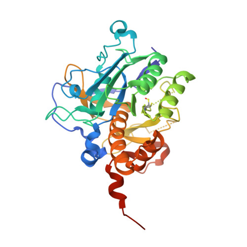
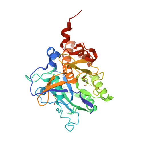
Crystal structure of an intracellular subtilisin reveals novel structural features unique to this subtilisin family.

Vevodova, J.Gamble, M.Kunze, G.Ariza, A.Dodson, E.Jones, D.D.Wilson, K.S.

(2010) Structure 18: 744-755

  • DOI: https://doi.org/10.1016/j.str.2010.03.008
  • Primary Citation of Related Structures:  
    2WV7, 2WWT, 2X8J

  • PubMed Abstract: 

    The intracellular subtilisin proteases (ISPs) are the only known members of the important and ubiquitous subtilisin family that function exclusively within the cell, constituting a major component of the degradome in many Gram-positive bacteria. The first ISP structure reported herein at a spacing of 1.56 A reveals features unique among subtilisins that has enabled potential functional and physiological roles to be assigned to sequence elements exclusive to the ISPs. Unlike all other subtilisins, ISP from B. clausii is dimeric, with residues from the C terminus making a major contribution to the dimer interface by crossing over to contact the partner subunit. A short N-terminal extension binds back across the active site to provide a potential novel regulatory mechanism of intrinsic proteolytic activity: a proline residue conserved throughout the ISPs introduces a kink in the polypeptide backbone that lifts the target peptide bond out of reach of the catalytic residues.


  • Organizational Affiliation

    Structural Biology Laboratory, Department of Chemistry, University of York, Heslington, York YO10 5YW, UK.


Macromolecules
Find similar proteins by:  (by identity cutoff)  |  3D Structure
Entity ID: 1
MoleculeChains Sequence LengthOrganismDetailsImage
INTRACELLULAR SUBTILISIN PROTEASE
A, C, D, E, F
327Shouchella clausiiMutation(s): 1 
EC: 3.4.21.62
UniProt
Find proteins for D0AB41 (Shouchella clausii)
Explore D0AB41 
Go to UniProtKB:  D0AB41
Entity Groups  
Sequence Clusters30% Identity50% Identity70% Identity90% Identity95% Identity100% Identity
UniProt GroupD0AB41
Sequence Annotations
Expand
  • Reference Sequence
Find similar proteins by:  (by identity cutoff)  |  3D Structure
Entity ID: 2
MoleculeChains Sequence LengthOrganismDetailsImage
INTRACELLULAR SUBTILISIN PROTEASE327Shouchella clausiiMutation(s): 1 
EC: 3.4.21.62
UniProt
Find proteins for D0AB41 (Shouchella clausii)
Explore D0AB41 
Go to UniProtKB:  D0AB41
Entity Groups  
Sequence Clusters30% Identity50% Identity70% Identity90% Identity95% Identity100% Identity
UniProt GroupD0AB41
Sequence Annotations
Expand
  • Reference Sequence
Small Molecules
Ligands 3 Unique
IDChains Name / Formula / InChI Key2D Diagram3D Interactions
1PE
Query on 1PE

Download Ideal Coordinates CCD File 
H [auth A]
J [auth B]
L [auth C]
O [auth D]
R [auth E]
H [auth A],
J [auth B],
L [auth C],
O [auth D],
R [auth E],
T [auth F]
PENTAETHYLENE GLYCOL
C10 H22 O6
JLFNLZLINWHATN-UHFFFAOYSA-N
GOL
Query on GOL

Download Ideal Coordinates CCD File 
M [auth C],
P [auth D]
GLYCEROL
C3 H8 O3
PEDCQBHIVMGVHV-UHFFFAOYSA-N
NA
Query on NA

Download Ideal Coordinates CCD File 
G [auth A]
I [auth B]
K [auth C]
N [auth D]
Q [auth E]
G [auth A],
I [auth B],
K [auth C],
N [auth D],
Q [auth E],
S [auth F]
SODIUM ION
Na
FKNQFGJONOIPTF-UHFFFAOYSA-N
Modified Residues  1 Unique
IDChains TypeFormula2D DiagramParent
CSX
Query on CSX
A, C, D, E, F
L-PEPTIDE LINKINGC3 H7 N O3 SCYS
Experimental Data & Validation

Experimental Data

  • Method: X-RAY DIFFRACTION
  • Resolution: 1.56 Å
  • R-Value Free: 0.230 
  • R-Value Work: 0.187 
  • R-Value Observed: 0.190 
  • Space Group: P 31
Unit Cell:
Length ( Å )Angle ( ˚ )
a = 125.659α = 90
b = 125.659β = 90
c = 106.141γ = 120
Software Package:
Software NamePurpose
REFMACrefinement
MOSFLMdata reduction
SCALAdata scaling
PHASERphasing

Structure Validation

View Full Validation Report


Entry History 

Deposition Data

Revision History  (Full details and data files)

  • Version 1.0: 2010-08-11
    Type: Initial release
  • Version 1.1: 2011-09-21
    Changes: Atomic model, Non-polymer description, Refinement description, Version format compliance
  • Version 1.2: 2018-02-28
    Changes: Advisory, Database references, Source and taxonomy
  • Version 1.3: 2019-09-25
    Changes: Data collection, Derived calculations, Experimental preparation, Other
  • Version 1.4: 2023-12-20
    Changes: Advisory, Data collection, Database references, Derived calculations, Refinement description