2WZS

Structure of the Family GH92 Inverting Mannosidase BT3990 from Bacteroides thetaiotaomicron VPI-5482 in complex with Mannoimidazole


Experimental Data Snapshot

  • Method: X-RAY DIFFRACTION
  • Resolution: 2.25 Å
  • R-Value Free: 0.242 
  • R-Value Work: 0.200 
  • R-Value Observed: 0.203 

wwPDB Validation   3D Report Full Report


Ligand Structure Quality Assessment 


This is version 1.2 of the entry. See complete history


Literature

Mechanistic Insights Into a Ca2+-Dependent Family of A-Mannosidases in a Human Gut Symbiont.

Zhu, Y.Suits, M.D.L.Thompson, A.Chavan, S.Dinev, Z.Dumon, C.Smith, N.Moremen, K.W.Xiang, Y.Siriwardena, A.Williams, S.J.Gilbert, H.J.Davies, G.J.

(2010) Nat Chem Biol 6: 125

  • DOI: https://doi.org/10.1038/nchembio.278
  • Primary Citation of Related Structures:  
    2WVX, 2WVY, 2WVZ, 2WW0, 2WW1, 2WW2, 2WW3, 2WZS

  • PubMed Abstract: 

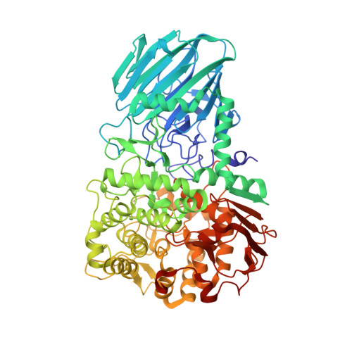
    Colonic bacteria, exemplified by Bacteroides thetaiotaomicron, play a key role in maintaining human health by harnessing large families of glycoside hydrolases (GHs) to exploit dietary polysaccharides and host glycans as nutrients. Such GH family expansion is exemplified by the 23 family GH92 glycosidases encoded by the B. thetaiotaomicron genome. Here we show that these are alpha-mannosidases that act via a single displacement mechanism to utilize host N-glycans. The three-dimensional structure of two GH92 mannosidases defines a family of two-domain proteins in which the catalytic center is located at the domain interface, providing acid (glutamate) and base (aspartate) assistance to hydrolysis in a Ca(2+)-dependent manner. The three-dimensional structures of the GH92s in complex with inhibitors provide insight into the specificity, mechanism and conformational itinerary of catalysis. Ca(2+) plays a key catalytic role in helping distort the mannoside away from its ground-state (4)C(1) chair conformation toward the transition state.


  • Organizational Affiliation

    Institute for Cell and Molecular Biosciences, The Medical School, Newcastle University, Framlington Place, Newcastle upon Tyne, UK.


Macromolecules
Find similar proteins by:  (by identity cutoff)  |  3D Structure
Entity ID: 1
MoleculeChains Sequence LengthOrganismDetailsImage
PUTATIVE ALPHA-1,2-MANNOSIDASE
A, B, C, D, E
A, B, C, D, E, F, G, H
738Bacteroides thetaiotaomicron VPI-5482Mutation(s): 0 
UniProt
Find proteins for Q8A0N1 (Bacteroides thetaiotaomicron (strain ATCC 29148 / DSM 2079 / JCM 5827 / CCUG 10774 / NCTC 10582 / VPI-5482 / E50))
Explore Q8A0N1 
Go to UniProtKB:  Q8A0N1
Entity Groups  
Sequence Clusters30% Identity50% Identity70% Identity90% Identity95% Identity100% Identity
UniProt GroupQ8A0N1
Sequence Annotations
Expand
  • Reference Sequence
Small Molecules
Ligands 3 Unique
IDChains Name / Formula / InChI Key2D Diagram3D Interactions
MVL
Query on MVL

Download Ideal Coordinates CCD File 
AA [auth D]
GA [auth E]
J [auth A]
LA [auth F]
O [auth B]
AA [auth D],
GA [auth E],
J [auth A],
LA [auth F],
O [auth B],
OA [auth G],
QA [auth H],
U [auth C]
(5R,6R,7S,8R)-5-(HYDROXYMETHYL)-5,6,7,8-TETRAHYDROIMIDAZO[1,2-A]PYRIDINE-6,7,8-TRIOL
C8 H12 N2 O4
RZRDQZQPTISYKY-JWXFUTCRSA-N
GOL
Query on GOL

Download Ideal Coordinates CCD File 
BA [auth D]
CA [auth D]
DA [auth D]
EA [auth D]
HA [auth E]
BA [auth D],
CA [auth D],
DA [auth D],
EA [auth D],
HA [auth E],
IA [auth E],
JA [auth E],
K [auth A],
L [auth A],
M [auth A],
MA [auth F],
P [auth B],
Q [auth B],
R [auth B],
S [auth B],
V [auth C],
W [auth C],
X [auth C],
Y [auth C]
GLYCEROL
C3 H8 O3
PEDCQBHIVMGVHV-UHFFFAOYSA-N
CA
Query on CA

Download Ideal Coordinates CCD File 
FA [auth E]
I [auth A]
KA [auth F]
N [auth B]
NA [auth G]
FA [auth E],
I [auth A],
KA [auth F],
N [auth B],
NA [auth G],
PA [auth H],
T [auth C],
Z [auth D]
CALCIUM ION
Ca
BHPQYMZQTOCNFJ-UHFFFAOYSA-N
Binding Affinity Annotations 
IDSourceBinding Affinity
MVL BindingDB:  2WZS Ki: 6.00e+5 (nM) from 1 assay(s)
Binding MOAD:  2WZS Ki: 1.40e+5 (nM) from 1 assay(s)
PDBBind:  2WZS Ki: 1.40e+5 (nM) from 1 assay(s)
Experimental Data & Validation

Experimental Data

  • Method: X-RAY DIFFRACTION
  • Resolution: 2.25 Å
  • R-Value Free: 0.242 
  • R-Value Work: 0.200 
  • R-Value Observed: 0.203 
  • Space Group: P 1 21 1
Unit Cell:
Length ( Å )Angle ( ˚ )
a = 109.19α = 90
b = 152.39β = 94.21
c = 221.45γ = 90
Software Package:
Software NamePurpose
REFMACrefinement
MOSFLMdata reduction
SCALAdata scaling
PHASERphasing

Structure Validation

View Full Validation Report


Ligand Structure Quality Assessment 


Entry History 

Revision History  (Full details and data files)

  • Version 1.0: 2009-12-29
    Type: Initial release
  • Version 1.1: 2011-05-08
    Changes: Version format compliance
  • Version 1.2: 2011-07-13
    Changes: Version format compliance