2WVS

Crystal structure of an alpha-L-fucosidase GH29 trapped covalent intermediate from Bacteroides thetaiotaomicron in complex with 2- fluoro-fucosyl fluoride using an E288Q mutant


Experimental Data Snapshot

  • Method: X-RAY DIFFRACTION
  • Resolution: 2.19 Å
  • R-Value Free: 0.235 
  • R-Value Work: 0.178 
  • R-Value Observed: 0.181 

wwPDB Validation   3D Report Full Report


Ligand Structure Quality Assessment 


This is version 1.3 of the entry. See complete history


Literature

Analysis of the Reaction Coordinate of Alpha-L-Fucosidases: A Combined Structural and Quantum Mechanical Approach

Lammerts Van Bueren, A.Ardevol, A.Fayers-Kerr, J.Luo, B.Zhang, Y.Sollogoub, M.Bleriot, Y.Rovira, C.Davies, G.J.

(2010) J Am Chem Soc 132: 1804

  • DOI: https://doi.org/10.1021/ja908908q
  • Primary Citation of Related Structures:  
    2WVS, 2WVT, 2WVU, 2WVV

  • PubMed Abstract: 

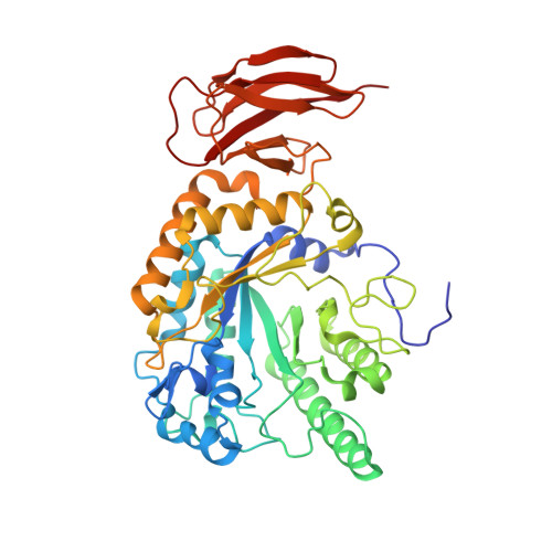
    The enzymatic hydrolysis of alpha-L-fucosides is of importance in cancer, bacterial infections, and fucosidosis, a neurodegenerative lysosomal storage disorder. Here we show a series of snapshots along the reaction coordinate of a glycoside hydrolase family GH29 alpha-L-fucosidase unveiling a Michaelis (ES) complex in a (1)C(4) (chair) conformation and a covalent glycosyl-enzyme intermediate in (3)S(1) (skew-boat). First principles metadynamics simulations on isolated alpha-L-fucose strongly support a (1)C(4)<-->(3)H(4)<-->(3)S(1) conformational itinerary for the glycosylation step of the reaction mechanism and indicate a strong "preactivation" of the (1)C(4) complex to nucleophilic attack as reflected by free energy, C1-O1/O5-C1 bond length elongation/reduction, C1-O1 bond orientation, and positive charge development around the anomeric carbon. Analysis of an imino sugar inhibitor is consistent with tight binding of a chair-conformed charged species.


  • Organizational Affiliation

    York Structural Biology Laboratory, Department of Chemistry, The University of York, York YO10 5YW, UK.


Macromolecules
Find similar proteins by:  (by identity cutoff)  |  3D Structure
Entity ID: 1
MoleculeChains Sequence LengthOrganismDetailsImage
ALPHA-L-FUCOSIDASE
A, B, C, D
443Bacteroides thetaiotaomicron VPI-5482Mutation(s): 1 
UniProt
Find proteins for Q8A3I4 (Bacteroides thetaiotaomicron (strain ATCC 29148 / DSM 2079 / JCM 5827 / CCUG 10774 / NCTC 10582 / VPI-5482 / E50))
Explore Q8A3I4 
Go to UniProtKB:  Q8A3I4
Entity Groups  
Sequence Clusters30% Identity50% Identity70% Identity90% Identity95% Identity100% Identity
UniProt GroupQ8A3I4
Sequence Annotations
Expand
  • Reference Sequence
Small Molecules
Ligands 2 Unique
IDChains Name / Formula / InChI Key2D Diagram3D Interactions
FUF
Query on FUF

Download Ideal Coordinates CCD File 
E [auth A],
F [auth A],
I [auth B],
K [auth C],
N [auth D]
2-deoxy-2-fluoro-beta-L-fucopyranose
C6 H11 F O4
IRKXGKIPOMIQOD-QYESYBIKSA-N
SO4
Query on SO4

Download Ideal Coordinates CCD File 
G [auth A]
H [auth A]
J [auth B]
L [auth C]
M [auth C]
G [auth A],
H [auth A],
J [auth B],
L [auth C],
M [auth C],
O [auth D],
P [auth D]
SULFATE ION
O4 S
QAOWNCQODCNURD-UHFFFAOYSA-L
Experimental Data & Validation

Experimental Data

  • Method: X-RAY DIFFRACTION
  • Resolution: 2.19 Å
  • R-Value Free: 0.235 
  • R-Value Work: 0.178 
  • R-Value Observed: 0.181 
  • Space Group: P 1 21 1
Unit Cell:
Length ( Å )Angle ( ˚ )
a = 54.84α = 90
b = 185.16β = 94.57
c = 97.87γ = 90
Software Package:
Software NamePurpose
REFMACrefinement

Structure Validation

View Full Validation Report


Ligand Structure Quality Assessment 


Entry History 

Deposition Data

Revision History  (Full details and data files)

  • Version 1.0: 2010-02-02
    Type: Initial release
  • Version 1.1: 2011-06-09
    Changes: Version format compliance
  • Version 1.2: 2011-07-13
    Changes: Version format compliance
  • Version 1.3: 2020-07-29
    Type: Remediation
    Reason: Carbohydrate remediation
    Changes: Data collection, Derived calculations, Other, Structure summary