2WTG

High resolution 3D structure of C.elegans globin-like protein GLB-1


Experimental Data Snapshot

  • Method: X-RAY DIFFRACTION
  • Resolution: 1.50 Å
  • R-Value Free: 0.201 
  • R-Value Work: 0.163 
  • R-Value Observed: 0.167 

wwPDB Validation   3D Report Full Report


Ligand Structure Quality Assessment 


This is version 1.2 of the entry. See complete history


Literature

Globin-Like Proteins in Caenorhabditis Elegans: In Vivo Localization, Ligand Binding and Structural Properties.

Geuens, E.Hoogewijs, D.Nardini, M.Vinck, E.Pesce, A.Kiger, L.Fago, A.Tilleman, L.De Henau, S.Marden, M.Weber, R.E.Van Doorslaer, S.Vanfleteren, J.Moens, L.Bolognesi, M.De Wilde, S.

(2010) BMC Biochem 11: 17

  • DOI: https://doi.org/10.1186/1471-2091-11-17
  • Primary Citation of Related Structures:  
    2WTG, 2WTH

  • PubMed Abstract: 

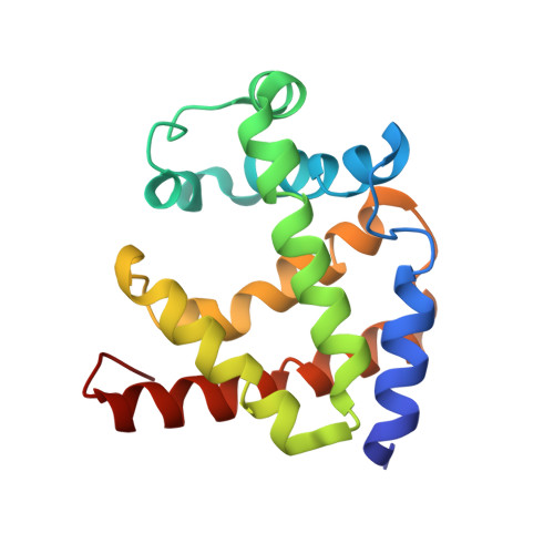
    The genome of the nematode Caenorhabditis elegans contains more than 30 putative globin genes that all are transcribed. Although their translated amino acid sequences fit the globin fold, a variety of amino-acid substitutions and extensions generate a wide structural diversity among the putative globins. No information is available on the physicochemical properties and the in vivo expression.


  • Organizational Affiliation

    Department of Biomedical Sciences, University of Antwerp, Universiteitsplein 1, B-2610 Antwerp, Belgium.


Macromolecules
Find similar proteins by:  (by identity cutoff)  |  3D Structure
Entity ID: 1
MoleculeChains Sequence LengthOrganismDetailsImage
GLOBIN-LIKE PROTEIN159Caenorhabditis elegansMutation(s): 1 
UniProt
Find proteins for P30627 (Caenorhabditis elegans)
Explore P30627 
Go to UniProtKB:  P30627
Entity Groups  
Sequence Clusters30% Identity50% Identity70% Identity90% Identity95% Identity100% Identity
UniProt GroupP30627
Sequence Annotations
Expand
  • Reference Sequence
Small Molecules
Ligands 2 Unique
IDChains Name / Formula / InChI Key2D Diagram3D Interactions
HEM
Query on HEM

Download Ideal Coordinates CCD File 
B [auth A]PROTOPORPHYRIN IX CONTAINING FE
C34 H32 Fe N4 O4
KABFMIBPWCXCRK-RGGAHWMASA-L
OXY
Query on OXY

Download Ideal Coordinates CCD File 
C [auth A]OXYGEN MOLECULE
O2
MYMOFIZGZYHOMD-UHFFFAOYSA-N
Experimental Data & Validation

Experimental Data

  • Method: X-RAY DIFFRACTION
  • Resolution: 1.50 Å
  • R-Value Free: 0.201 
  • R-Value Work: 0.163 
  • R-Value Observed: 0.167 
  • Space Group: P 43 21 2
Unit Cell:
Length ( Å )Angle ( ˚ )
a = 81.947α = 90
b = 81.947β = 90
c = 47.042γ = 90
Software Package:
Software NamePurpose
REFMACrefinement
MOSFLMdata reduction
SCALAdata scaling
SOLVEphasing

Structure Validation

View Full Validation Report


Ligand Structure Quality Assessment 


Entry History 

Revision History  (Full details and data files)

  • Version 1.0: 2010-04-21
    Type: Initial release
  • Version 1.1: 2011-05-08
    Changes: Version format compliance
  • Version 1.2: 2011-07-13
    Changes: Version format compliance