The Crystal Structures of Macrophage Migration Inhibition Factor (Mif) from Plasmodium Falciparum and Plasmodium Berghei.
Dobson, S.E., Augustijn, K.D., Brannigan, J.A., Schnick, C., Janse, C.J., Dodson, E.J., Waters, A.P., Wilkinson, A.J.(2009) Protein Sci 18: 2578
- PubMed: 19827093
- DOI: https://doi.org/10.1002/pro.263
- Primary Citation of Related Structures:
2WKB, 2WKF - PubMed Abstract:
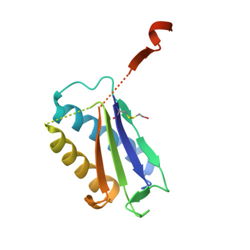
Malaria, caused by Plasmodium falciparum and related parasites, is responsible for millions of deaths each year, mainly from complications arising from the blood stages of its life cycle. Macrophage migration inhibitory factor (MIF), a protein expressed by the parasite during these stages, has been characterized in mammals as a cytokine involved in a broad spectrum of immune responses. It also possesses two catalytic activities, a tautomerase and an oxidoreductase, though the physiological significance of neither reaction is known. Here, we have determined the crystal structure of MIF from two malaria parasites, Plasmodium falciparum and Plasmodium berghei at 2.2 A and 1.8 A, respectively. The structures have an alpha/beta fold and each reveals a trimer, in agreement with the results of analytical ultracentrifugation. We observed open and closed active sites, these being distinguished by movements of proline-1, the catalytic base in the tautomerase reaction. These states correlate with the covalent modification of cysteine 2 to form a mercaptoethanol adduct, an observation confirmed by mass spectrometry. The Plasmodium MIFs have a different pattern of conserved cysteine residues to the mammalian MIFs and the side chain of Cys58, which is implicated in the oxidoreductase activity, is buried. This observation and the evident redox reactivity of Cys2 suggest quite different oxidoreductase characteristics. Finally, we show in pull-down assays that Plasmodium MIF binds to the cell surface receptor CD74, a known mammalian MIF receptor implying that parasite MIF has the ability to interfere with, or modulate, host MIF activity through a competitive binding mechanism.
Organizational Affiliation:
Structural Biology Laboratory, Department of Chemistry, University of York, York YO10 5YW, United Kingdom.