2VZK

Structure of the acyl-enzyme complex of an N-terminal nucleophile (Ntn) hydrolase, OAT2


Experimental Data Snapshot

  • Method: X-RAY DIFFRACTION
  • Resolution: 2.33 Å
  • R-Value Free: 0.284 
  • R-Value Work: 0.254 
  • R-Value Observed: 0.255 

wwPDB Validation   3D Report Full Report


This is version 3.1 of the entry. See complete history


Literature

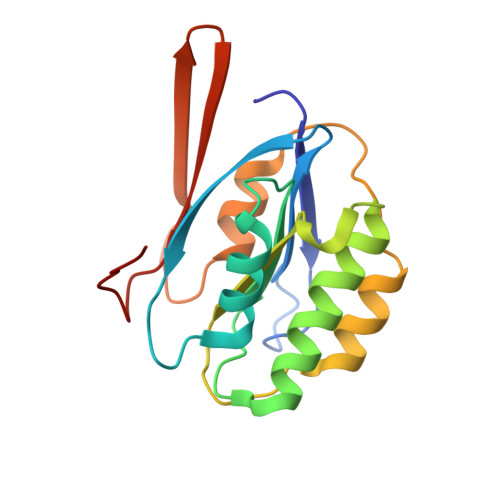
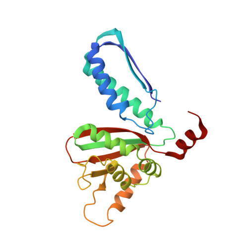
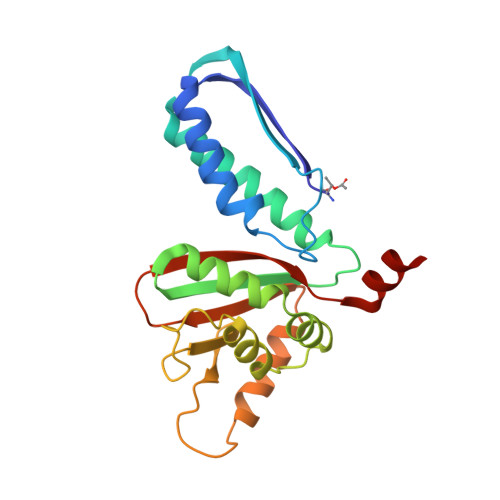
Anatomy of a Simple Acyl Intermediate in Enzyme Catalysis: Combined Biophysical and Modeling Studies on Ornithine Acetyl Transferase.

Iqbal, A.Clifton, I.J.Bagonis, M.Kershaw, N.J.Domene, C.Claridge, T.D.Wharton, C.W.Schofield, C.J.

(2009) J Am Chem Soc 131: 749

  • DOI: https://doi.org/10.1021/ja807215u
  • Primary Citation of Related Structures:  
    2VZK

  • PubMed Abstract: 

    Acyl-enzyme complexes are intermediates in reactions catalyzed by many hydrolases and related enzymes which employ nucleophilic catalysis. However, most of the reported structural data on acyl-enzyme complexes has been acquired under noncatalytic conditions. Recent IR analyses have indicated that some acyl-enzyme complexes may be more flexible than most crystallographic analyses have implied. OAT2 is a member of the N-terminal nucleophile (Ntn) hydrolase enzyme superfamily and catalyzes the reversible transfer of an acetyl group between the alpha-amino groups of ornithine and glutamate in a mechanism proposed to involve an acyl-enzyme complex. We have carried out biophysical analyses on ornithine acetyl transferase (OAT2), both in solution and in the crystalline state. Mass spectrometric studies identified Thr-181 as the residue acetylated during OAT2 catalysis; (13)C NMR analyses implied the presence of an acyl-enzyme complex in solution. Crystallization of OAT2 in the presence of N-alpha-acetyl-L-glutamate led to a structure in which Thr-181 was acetylated; the carbonyl oxygen of the acyl-enzyme complex was located in an oxyanion hole and positioned to hydrogen bond with the backbone amide NH of Gly-112 and the alcohol of Thr-111. While the crystallographic analyses revealed only one structure, IR spectroscopy demonstrated the presence of two distinct acyl-enzyme complex structures with carbonyl stretching frequencies at 1691 and 1701 cm(-1). Modeling studies implied two possible acyl-enzyme complex structures, one of which correlates with that observed in the crystal structure and with the 1691 cm(-1) IR absorption. The second acyl-enzyme complex structure, which has only a single oxyanion hole hydrogen bond, is proposed to give rise to the 1701 cm(-1) IR absorption. The two acyl-enzyme complex structures can interconvert by movement of the Thr-111 side-chain alcohol hydrogen away from the oxyanion hole to hydrogen bond with the backbone carbonyl of the acylated residue, Thr-181. Overall, the results reveal that acyl-enzyme complex structures may be more dynamic than previously thought and support the use of a comprehensive biophysical and modeling approach in studying such intermediates.


  • Organizational Affiliation

    Chemistry Research Laboratory, University of Oxford, 12 Mansfield Road, Oxford, OX1 3TA, UK.


Macromolecules
Find similar proteins by:  (by identity cutoff)  |  3D Structure
Entity ID: 1
MoleculeChains Sequence LengthOrganismDetailsImage
GLUTAMATE N-ACETYLTRANSFERASE 2 ALPHA CHAIN
A, C, E, G
173Streptomyces clavuligerusMutation(s): 0 
EC: 2.3.1.35
UniProt
Find proteins for P0DJQ5 (Streptomyces clavuligerus)
Explore P0DJQ5 
Go to UniProtKB:  P0DJQ5
Entity Groups  
Sequence Clusters30% Identity50% Identity70% Identity90% Identity95% Identity100% Identity
UniProt GroupP0DJQ5
Sequence Annotations
Expand
  • Reference Sequence
Find similar proteins by:  (by identity cutoff)  |  3D Structure
Entity ID: 2
MoleculeChains Sequence LengthOrganismDetailsImage
GLUTAMATE N-ACETYLTRANSFERASE 2 BETA CHAIN213Streptomyces clavuligerusMutation(s): 0 
EC: 2.3.1.35
UniProt
Find proteins for P0DJQ5 (Streptomyces clavuligerus)
Explore P0DJQ5 
Go to UniProtKB:  P0DJQ5
Entity Groups  
Sequence Clusters30% Identity50% Identity70% Identity90% Identity95% Identity100% Identity
UniProt GroupP0DJQ5
Sequence Annotations
Expand
  • Reference Sequence
Find similar proteins by:  (by identity cutoff)  |  3D Structure
Entity ID: 3
MoleculeChains Sequence LengthOrganismDetailsImage
GLUTAMATE N-ACETYLTRANSFERASE 2 BETA CHAIN
D, F, H
213Streptomyces clavuligerusMutation(s): 0 
EC: 2.3.1.35
UniProt
Find proteins for P0DJQ5 (Streptomyces clavuligerus)
Explore P0DJQ5 
Go to UniProtKB:  P0DJQ5
Entity Groups  
Sequence Clusters30% Identity50% Identity70% Identity90% Identity95% Identity100% Identity
UniProt GroupP0DJQ5
Sequence Annotations
Expand
  • Reference Sequence
Small Molecules
Experimental Data & Validation

Experimental Data

  • Method: X-RAY DIFFRACTION
  • Resolution: 2.33 Å
  • R-Value Free: 0.284 
  • R-Value Work: 0.254 
  • R-Value Observed: 0.255 
  • Space Group: P 1 21 1
Unit Cell:
Length ( Å )Angle ( ˚ )
a = 61.273α = 90
b = 73.878β = 92.71
c = 172.166γ = 90
Software Package:
Software NamePurpose
REFMACrefinement
HKL-2000data reduction
SCALEPACKdata scaling
PHASERphasing

Structure Validation

View Full Validation Report


Entry History 

Deposition Data

Revision History  (Full details and data files)

  • Version 1.0: 2008-09-16
    Type: Initial release
  • Version 1.1: 2011-05-07
    Changes: Version format compliance
  • Version 1.2: 2011-07-13
    Changes: Version format compliance
  • Version 1.3: 2017-07-05
    Changes: Data collection
  • Version 2.0: 2019-05-15
    Changes: Advisory, Data collection, Derived calculations, Experimental preparation, Polymer sequence
  • Version 3.0: 2023-11-15
    Changes: Atomic model, Data collection, Database references, Derived calculations, Other
  • Version 3.1: 2023-12-13
    Changes: Refinement description