2VT0

X-ray structure of a conjugate with conduritol-beta-epoxide of acid-beta-glucosidase overexpressed in cultured plant cells


Experimental Data Snapshot

  • Method: X-RAY DIFFRACTION
  • Resolution: 2.15 Å
  • R-Value Free: 0.201 
  • R-Value Work: 0.152 
  • R-Value Observed: 0.154 

wwPDB Validation   3D Report Full Report


Ligand Structure Quality Assessment 


This is version 2.1 of the entry. See complete history


Literature

Acid Beta-Glucosidase: Insights from Structural Analysis and Relevance to Gaucher Disease Therapy.

Kacher, Y.Brumshtein, B.Boldin-Adamsky, S.Toker, L.Shainskaya, A.Silman, I.Sussman, J.L.Futerman, A.H.

(2008) Biol Chem 389: 1361

  • DOI: https://doi.org/10.1515/BC.2008.163
  • Primary Citation of Related Structures:  
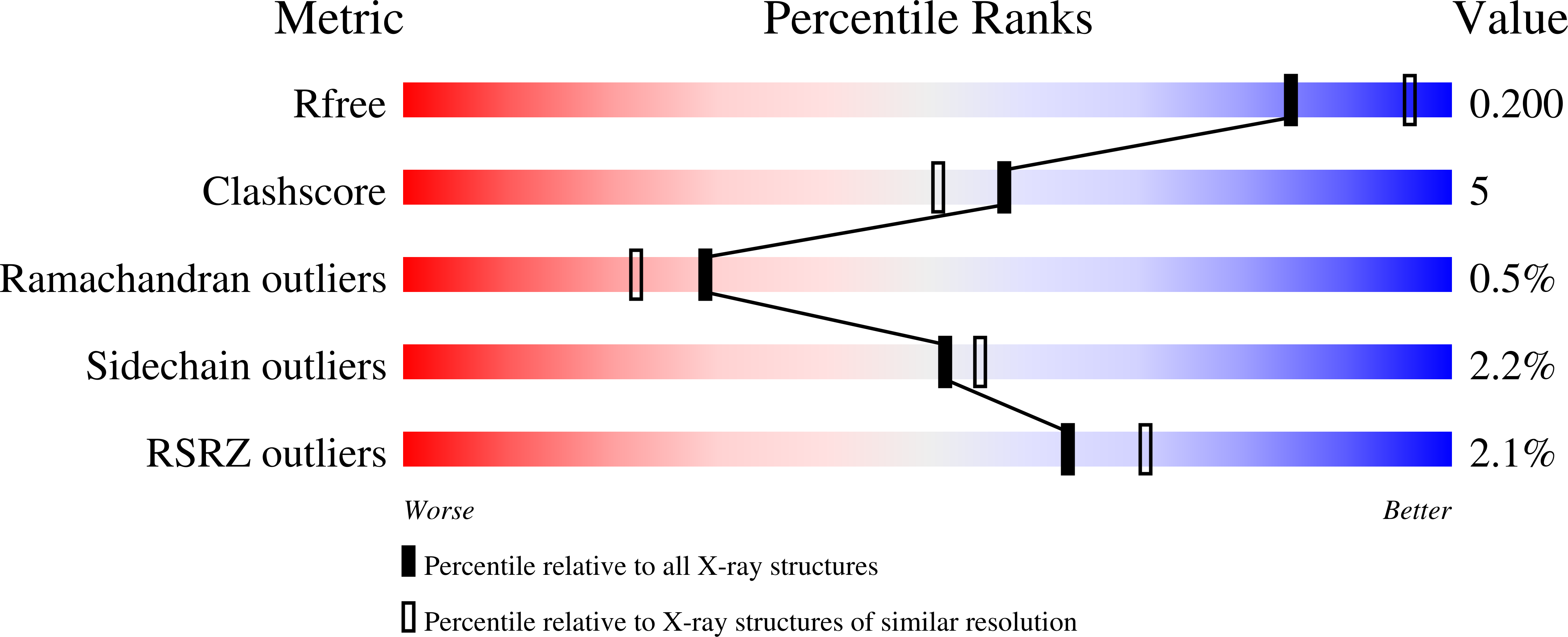
    2VT0

  • PubMed Abstract: 

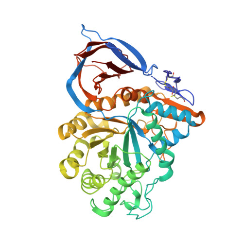
    In mammalian cells, glucosylceramide (GlcCer), the simplest glycosphingolipid, is hydrolyzed by the lysosomal enzyme acid beta-glucosidase (GlcCerase). In the human metabolic disorder Gaucher disease, GlcCerase activity is significantly decreased owing to one of approximately 200 mutations in the GlcCerase gene. The most common therapy for Gaucher disease is enzyme replacement therapy (ERT), in which patients are given intravenous injections of recombinant human GlcCerase; the Genzyme product Cerezyme has been used clinically for more than 15 years and is administered to approximately 4000 patients worldwide. Here we review the crystal structure of Cerezyme and other recombinant forms of GlcCerase, as well as of their complexes with covalent and non-covalent inhibitors. We also discuss the stability of Cerezyme, which can be altered by modification of its N-glycan chains with possible implications for improved ERT in Gaucher disease.


  • Organizational Affiliation

    Department of Biological Chemistry, Weizmann Institute of Science, Rehovot 76100, Israel.


Macromolecules
Find similar proteins by:  (by identity cutoff)  |  3D Structure
Entity ID: 1
MoleculeChains Sequence LengthOrganismDetailsImage
GLUCOSYLCERAMIDASE
A, B
505Homo sapiensMutation(s): 0 
EC: 3.2.1.45
Membrane Entity: Yes 
UniProt & NIH Common Fund Data Resources
Find proteins for P04062 (Homo sapiens)
Explore P04062 
Go to UniProtKB:  P04062
PHAROS:  P04062
GTEx:  ENSG00000177628 
Entity Groups  
Sequence Clusters30% Identity50% Identity70% Identity90% Identity95% Identity100% Identity
UniProt GroupP04062
Sequence Annotations
Expand
  • Reference Sequence
Oligosaccharides

Help

Entity ID: 2
MoleculeChains Length2D Diagram Glycosylation3D Interactions
alpha-D-mannopyranose-(1-4)-2-acetamido-2-deoxy-beta-D-glucopyranose-(1-4)-[alpha-L-fucopyranose-(1-3)]2-acetamido-2-deoxy-beta-D-glucopyranose
C
4N-Glycosylation
Glycosylation Resources
GlyTouCan:  G73622RM
GlyCosmos:  G73622RM
GlyGen:  G73622RM
Entity ID: 3
MoleculeChains Length2D Diagram Glycosylation3D Interactions
alpha-L-fucopyranose-(1-3)-[2-acetamido-2-deoxy-beta-D-glucopyranose-(1-4)]2-acetamido-2-deoxy-beta-D-glucopyranose
D
3N-Glycosylation
Glycosylation Resources
GlyTouCan:  G11971MR
GlyCosmos:  G11971MR
GlyGen:  G11971MR
Small Molecules
Ligands 2 Unique
IDChains Name / Formula / InChI Key2D Diagram3D Interactions
CBU
Query on CBU

Download Ideal Coordinates CCD File 
E [auth A],
L [auth B]
(1R,2R,3S,4S,5S,6S)-CYCLOHEXANE-1,2,3,4,5,6-HEXOL
C6 H12 O6
CDAISMWEOUEBRE-LKPKBOIGSA-N
SO4
Query on SO4

Download Ideal Coordinates CCD File 
F [auth A]
G [auth A]
H [auth A]
I [auth A]
J [auth A]
F [auth A],
G [auth A],
H [auth A],
I [auth A],
J [auth A],
K [auth A],
M [auth B],
N [auth B],
O [auth B],
P [auth B]
SULFATE ION
O4 S
QAOWNCQODCNURD-UHFFFAOYSA-L
Experimental Data & Validation

Experimental Data

  • Method: X-RAY DIFFRACTION
  • Resolution: 2.15 Å
  • R-Value Free: 0.201 
  • R-Value Work: 0.152 
  • R-Value Observed: 0.154 
  • Space Group: P 1 21 1
Unit Cell:
Length ( Å )Angle ( ˚ )
a = 68.184α = 90
b = 96.884β = 103.73
c = 83.171γ = 90
Software Package:
Software NamePurpose
REFMACrefinement
HKL-2000data reduction
HKL-2000data scaling
PHASERphasing

Structure Validation

View Full Validation Report


Ligand Structure Quality Assessment 


Entry History 

Deposition Data

Revision History  (Full details and data files)

  • Version 1.0: 2008-09-23
    Type: Initial release
  • Version 1.1: 2012-10-10
    Changes: Database references, Derived calculations, Non-polymer description, Other, Version format compliance
  • Version 1.2: 2019-05-08
    Changes: Data collection, Derived calculations, Experimental preparation, Other
  • Version 2.0: 2020-07-29
    Type: Remediation
    Reason: Carbohydrate remediation
    Changes: Advisory, Atomic model, Data collection, Derived calculations, Other, Structure summary
  • Version 2.1: 2023-12-13
    Changes: Data collection, Database references, Derived calculations, Refinement description, Structure summary