2VNV

Crystal structure of BclA lectin from burkholderia cenocepacia in complex with alpha-methyl-mannoside at 1.7 Angstrom resolution


Experimental Data Snapshot

  • Method: X-RAY DIFFRACTION
  • Resolution: 1.70 Å
  • R-Value Free: 0.203 
  • R-Value Work: 0.169 
  • R-Value Observed: 0.171 

wwPDB Validation   3D Report Full Report


Ligand Structure Quality Assessment 


This is version 1.3 of the entry. See complete history


Literature

Structural Basis for Mannose Recognition by a Lectin from Opportunistic Bacteria Burkholderia Cenocepacia

Lameignere, E.Malinovska, L.Slavikova, M.Duchaud, E.Mitchell, E.P.Varrot, A.Sedo, O.Imberty, A.Wimmerova, M.

(2008) Biochem J 411: 307

  • DOI: https://doi.org/10.1042/bj20071276
  • Primary Citation of Related Structures:  
    2VNV

  • PubMed Abstract: 

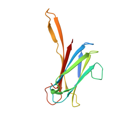
    Chronic colonization of the lungs by opportunist bacteria such as Pseudomonas aeruginosa and members of the Bcc (Burkholderia cepacia complex) is the major cause of morbidity and mortality among CF (cystic fibrosis) patients. PA-IIL (lecB gene), a soluble lectin from Ps. aeruginosa, has been the subject of much interest because of its very strong affinity for fucose. Orthologues have been identified in the opportunist bacteria Ralstonia solanacearum, Chromobacterium violaceum and Burkholderia of Bcc. The genome of the J2315 strain of B. cenocepacia, responsible for epidemia in CF centres, contains three genes that code for proteins with PA-IIL domains. The shortest gene was cloned in Escherichia coli and pure recombinant protein, BclA (B. cenocepacia lectin A), was obtained. The presence of native BclA in B. cenocepacia extracts was checked using a proteomic approach. The specificity of recombinant BclA was characterized using surface plasmon resonance showing a preference for mannosides and supported with glycan array experiments demonstrating a strict specificity for oligomannose-type N-glycan structures. The interaction thermodynamics of BclA with methyl alpha-D-mannoside demonstrates a dissociation constant (K(d)) of 2.75 x 10(-6) M. The X-ray crystal structure of the complex with methyl alpha-D-mannoside was determined at 1.7 A (1 A=0.1 nm) resolution. The lectin forms homodimers with one binding site per monomer, acting co-operatively with the second dimer site. Each monomer contains two Ca2+ ions and one sugar ligand. Despite strong sequence similarity, the differences between BclA and PA-IIL in their specificity, binding site and oligomerization mode indicate that the proteins should have different roles in the bacteria.


  • Organizational Affiliation

    CERMAV-CNRS, BP 53, F-38041, Grenoble, Cedex 09, France.


Macromolecules
Find similar proteins by:  (by identity cutoff)  |  3D Structure
Entity ID: 1
MoleculeChains Sequence LengthOrganismDetailsImage
BCLA
A, B, C, D, E
129Burkholderia cenocepacia J2315Mutation(s): 0 
UniProt
Find proteins for B4EH87 (Burkholderia cenocepacia (strain ATCC BAA-245 / DSM 16553 / LMG 16656 / NCTC 13227 / J2315 / CF5610))
Explore B4EH87 
Go to UniProtKB:  B4EH87
Entity Groups  
Sequence Clusters30% Identity50% Identity70% Identity90% Identity95% Identity100% Identity
UniProt GroupB4EH87
Sequence Annotations
Expand
  • Reference Sequence
Small Molecules
Ligands 3 Unique
IDChains Name / Formula / InChI Key2D Diagram3D Interactions
MMA
Query on MMA

Download Ideal Coordinates CCD File 
F [auth A],
J [auth B],
N [auth C],
Q [auth D],
U [auth E]
methyl alpha-D-mannopyranoside
C7 H14 O6
HOVAGTYPODGVJG-VEIUFWFVSA-N
SO4
Query on SO4

Download Ideal Coordinates CCD File 
I [auth A],
M [auth B],
T [auth D],
X [auth E]
SULFATE ION
O4 S
QAOWNCQODCNURD-UHFFFAOYSA-L
CA
Query on CA

Download Ideal Coordinates CCD File 
G [auth A]
H [auth A]
K [auth B]
L [auth B]
O [auth C]
G [auth A],
H [auth A],
K [auth B],
L [auth B],
O [auth C],
P [auth C],
R [auth D],
S [auth D],
V [auth E],
W [auth E]
CALCIUM ION
Ca
BHPQYMZQTOCNFJ-UHFFFAOYSA-N
Binding Affinity Annotations 
IDSourceBinding Affinity
MMA Binding MOAD:  2VNV Kd: 2750 (nM) from 1 assay(s)
Experimental Data & Validation

Experimental Data

  • Method: X-RAY DIFFRACTION
  • Resolution: 1.70 Å
  • R-Value Free: 0.203 
  • R-Value Work: 0.169 
  • R-Value Observed: 0.171 
  • Space Group: C 2 2 21
Unit Cell:
Length ( Å )Angle ( ˚ )
a = 50.01α = 90
b = 185.702β = 90
c = 187.551γ = 90
Software Package:
Software NamePurpose
REFMACrefinement
MOSFLMdata reduction
SCALAdata scaling
MOLREPphasing

Structure Validation

View Full Validation Report


Ligand Structure Quality Assessment 


Entry History 

Deposition Data

Revision History  (Full details and data files)

  • Version 1.0: 2008-04-08
    Type: Initial release
  • Version 1.1: 2011-05-10
    Changes: Version format compliance
  • Version 1.2: 2011-07-13
    Changes: Version format compliance
  • Version 1.3: 2020-07-29
    Type: Remediation
    Reason: Carbohydrate remediation
    Changes: Data collection, Derived calculations, Other, Structure summary