2VK3

Crystal structure of rat profilin 2a


Experimental Data Snapshot

  • Method: X-RAY DIFFRACTION
  • Resolution: 1.70 Å
  • R-Value Free: 0.216 
  • R-Value Work: 0.173 
  • R-Value Observed: 0.175 

wwPDB Validation   3D Report Full Report


Ligand Structure Quality Assessment 


This is version 1.5 of the entry. See complete history


Literature

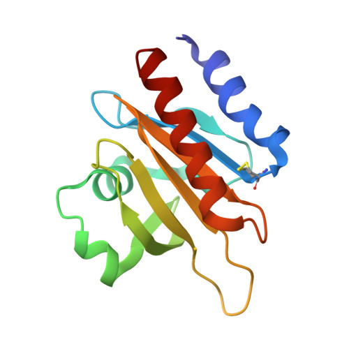
Structure, Modifications and Ligand-Binding Properties of Rat Profilin 2A.

Haikarainen, T.Chen, W.Q.Lubec, G.Kursula, P.

(2009) Acta Crystallogr D Biol Crystallogr 65: 303

  • DOI: https://doi.org/10.1107/S0907444909000699
  • Primary Citation of Related Structures:  
    2VK3

  • PubMed Abstract: 

    Profilins are key regulators of the actin microfilament system and in neuronal tissues the profilin 2a isoform is the most abundant and important profilin. The high-resolution crystal structure of rat profilin 2a has been determined in the absence of ligands. By comparing the structure with those of peptide-liganded profilin 2a and unliganded profilin 2b, it can be concluded that the binding site for proline-rich peptides is pre-organized. The C-terminus of profilin 2a is also well ordered in the absence of ligand peptide, in contrast to the 2b isoform which is generated by alternative splicing. Covalent modifications of four cysteine residues were also detected in profilin 2a, as well as a number of other modifications in profilin 2 from rat brain; such modifications could significantly affect the function of profilin. It was also shown that profilin 2a binds to the neuronal protein palladin, including a synthetic palladin peptide; peptides from another profilin ligand, dynamin 1, failed to interact with both profilin 1 and profilin 2a. These results allow a better understanding of the structure-function relationships and ligand binding of mammalian profilin 2a.


  • Organizational Affiliation

    Department of Biochemistry, University of Oulu, Oulu, Finland.


Macromolecules
Find similar proteins by:  (by identity cutoff)  |  3D Structure
Entity ID: 1
MoleculeChains Sequence LengthOrganismDetailsImage
PROFILIN-2142Rattus norvegicusMutation(s): 0 
UniProt
Find proteins for Q9EPC6 (Rattus norvegicus)
Explore Q9EPC6 
Go to UniProtKB:  Q9EPC6
Entity Groups  
Sequence Clusters30% Identity50% Identity70% Identity90% Identity95% Identity100% Identity
UniProt GroupQ9EPC6
Sequence Annotations
Expand
  • Reference Sequence
Experimental Data & Validation

Experimental Data

  • Method: X-RAY DIFFRACTION
  • Resolution: 1.70 Å
  • R-Value Free: 0.216 
  • R-Value Work: 0.173 
  • R-Value Observed: 0.175 
  • Space Group: P 41
Unit Cell:
Length ( Å )Angle ( ˚ )
a = 51.1α = 90
b = 51.1β = 90
c = 67.52γ = 90
Software Package:
Software NamePurpose
REFMACrefinement
XDSdata reduction
XSCALEdata scaling
MOLREPphasing

Structure Validation

View Full Validation Report


Ligand Structure Quality Assessment 


Entry History 

Deposition Data

Revision History  (Full details and data files)

  • Version 1.0: 2009-02-10
    Type: Initial release
  • Version 1.1: 2011-05-08
    Changes: Version format compliance
  • Version 1.2: 2011-07-13
    Changes: Version format compliance
  • Version 1.3: 2018-03-07
    Changes: Source and taxonomy
  • Version 1.4: 2019-07-24
    Changes: Data collection, Derived calculations
  • Version 1.5: 2023-12-13
    Changes: Data collection, Database references, Derived calculations, Other, Refinement description