2VK2

Crystal structure of a galactofuranose binding protein


Experimental Data Snapshot

  • Method: X-RAY DIFFRACTION
  • Resolution: 1.20 Å
  • R-Value Free: 0.190 
  • R-Value Work: 0.178 
  • R-Value Observed: 0.179 

wwPDB Validation   3D Report Full Report


Ligand Structure Quality Assessment 


This is version 2.1 of the entry. See complete history


Literature

Furanose-Specific Sugar Transport: Characterization of a Bacterial Galactofuranose-Binding Protein.

Horler, R.S.P.Muller, A.Williamson, D.C.Potts, J.R.Wilson, K.S.Thomas, G.H.

(2009) J Biol Chem 284: 31156

  • DOI: https://doi.org/10.1074/jbc.M109.054296
  • Primary Citation of Related Structures:  
    2VK2

  • PubMed Abstract: 

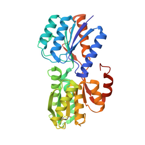
    The widespread utilization of sugars by microbes is reflected in the diversity and multiplicity of cellular transporters used to acquire these compounds from the environment. The model bacterium Escherichia coli has numerous transporters that allow it to take up hexoses and pentoses, which recognize the more abundant pyranose forms of these sugars. Here we report the biochemical and structural characterization of a transporter protein YtfQ from E. coli that forms part of an uncharacterized ABC transporter system. Remarkably the crystal structure of this protein, solved to 1.2 A using x-ray crystallography, revealed that YtfQ binds a single molecule of galactofuranose in its ligand binding pocket. Selective binding of galactofuranose over galactopyranose was also observed using NMR methods that determined the form of the sugar released from the protein. The pattern of expression of the ytfQRTyjfF operon encoding this transporter mirrors that of the high affinity galactopyranose transporter of E. coli, suggesting that this bacterium has evolved complementary transporters that enable it to use all the available galactose present during carbon limiting conditions.


  • Organizational Affiliation

    Department of Biology, University of York, York YO10 5YW, United Kingdom.


Macromolecules
Find similar proteins by:  (by identity cutoff)  |  3D Structure
Entity ID: 1
MoleculeChains Sequence LengthOrganismDetailsImage
ABC TRANSPORTER PERIPLASMIC-BINDING PROTEIN YTFQ306Escherichia coli K-12Mutation(s): 0 
UniProt
Find proteins for P39325 (Escherichia coli (strain K12))
Explore P39325 
Go to UniProtKB:  P39325
Entity Groups  
Sequence Clusters30% Identity50% Identity70% Identity90% Identity95% Identity100% Identity
UniProt GroupP39325
Sequence Annotations
Expand
  • Reference Sequence
Small Molecules
Ligands 1 Unique
IDChains Name / Formula / InChI Key2D Diagram3D Interactions
GZL
Query on GZL

Download Ideal Coordinates CCD File 
B [auth A]beta-D-galactofuranose
C6 H12 O6
AVVWPBAENSWJCB-DGPNFKTASA-N
Binding Affinity Annotations 
IDSourceBinding Affinity
GZL PDBBind:  2VK2 Kd: 130 (nM) from 1 assay(s)
Binding MOAD:  2VK2 Kd: 130 (nM) from 1 assay(s)
Experimental Data & Validation

Experimental Data

  • Method: X-RAY DIFFRACTION
  • Resolution: 1.20 Å
  • R-Value Free: 0.190 
  • R-Value Work: 0.178 
  • R-Value Observed: 0.179 
  • Space Group: P 21 21 21
Unit Cell:
Length ( Å )Angle ( ˚ )
a = 45.745α = 90
b = 51.535β = 90
c = 116.091γ = 90
Software Package:
Software NamePurpose
REFMACrefinement
DENZOdata reduction
SCALEPACKdata scaling
PHASERphasing

Structure Validation

View Full Validation Report


Ligand Structure Quality Assessment 


Entry History 

Deposition Data

Revision History  (Full details and data files)

  • Version 1.0: 2009-05-19
    Type: Initial release
  • Version 1.1: 2011-05-08
    Changes: Version format compliance
  • Version 1.2: 2011-07-13
    Changes: Version format compliance
  • Version 2.0: 2020-07-29
    Type: Remediation
    Reason: Carbohydrate remediation
    Changes: Advisory, Atomic model, Data collection, Database references, Derived calculations, Other, Structure summary
  • Version 2.1: 2023-12-13
    Changes: Data collection, Database references, Refinement description, Structure summary