2V7G

Crystal Structure of an Engineered Urocanase Tetramer


Experimental Data Snapshot

  • Method: X-RAY DIFFRACTION
  • Resolution: 2.00 Å
  • R-Value Free: 0.181 
  • R-Value Work: 0.156 
  • R-Value Observed: 0.156 

wwPDB Validation   3D Report Full Report


Ligand Structure Quality Assessment 


This is version 1.3 of the entry. See complete history


Literature

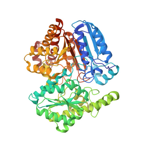
Designed Protein-Protein Association.

Grueninger, D.Treiber, N.Ziegler, M.O.P.Koetter, J.W.A.Schulze, M.-S.Schulz, G.E.

(2008) Science 319: 206

  • DOI: https://doi.org/10.1126/science.1150421
  • Primary Citation of Related Structures:  
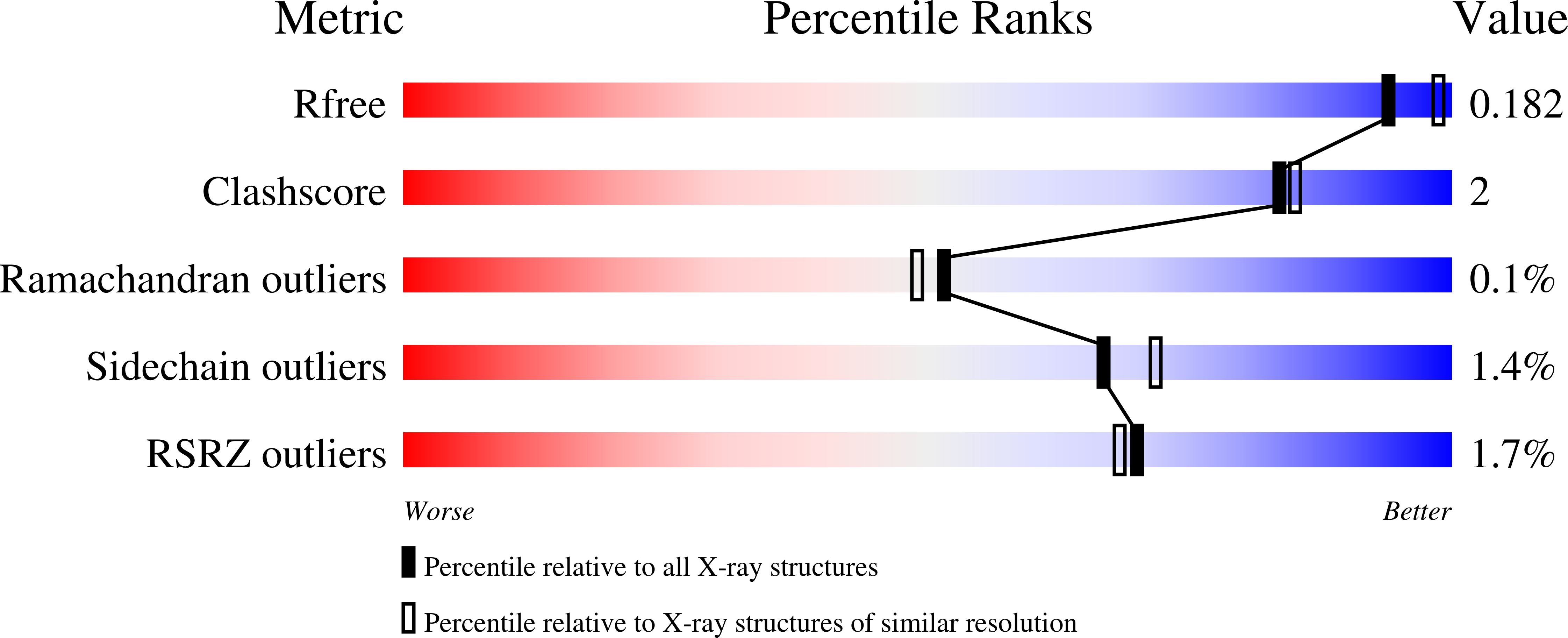
    2UYU, 2UYV, 2V7G, 2V9E, 2V9F, 2V9G, 2V9I, 2V9L, 2V9M, 2V9N, 2V9O, 2V9U

  • PubMed Abstract: 

    The analysis of natural contact interfaces between protein subunits and between proteins has disclosed some general rules governing their association. We have applied these rules to produce a number of novel assemblies, demonstrating that a given protein can be engineered to form contacts at various points of its surface. Symmetry plays an important role because it defines the multiplicity of a designed contact and therefore the number of required mutations. Some of the proteins needed only a single side-chain alteration in order to associate to a higher-order complex. The mobility of the buried side chains has to be taken into account. Four assemblies have been structurally elucidated. Comparisons between the designed contacts and the results will provide useful guidelines for the development of future architectures.


  • Organizational Affiliation

    Institut für Organische Chemie und Biochemie, Albert-Ludwigs-Universität, Albertstrasse 21, 79104 Freiburg im Breisgau, Germany.


Macromolecules
Find similar proteins by:  (by identity cutoff)  |  3D Structure
Entity ID: 1
MoleculeChains Sequence LengthOrganismDetailsImage
UROCANATE HYDRATASE
A, B, C, D
557Pseudomonas putidaMutation(s): 4 
EC: 4.2.1.49
UniProt
Find proteins for P25080 (Pseudomonas putida)
Explore P25080 
Go to UniProtKB:  P25080
Entity Groups  
Sequence Clusters30% Identity50% Identity70% Identity90% Identity95% Identity100% Identity
UniProt GroupP25080
Sequence Annotations
Expand
  • Reference Sequence
Small Molecules
Ligands 3 Unique
IDChains Name / Formula / InChI Key2D Diagram3D Interactions
NAD
Query on NAD

Download Ideal Coordinates CCD File 
I [auth A],
P [auth B],
S [auth C],
X [auth D]
NICOTINAMIDE-ADENINE-DINUCLEOTIDE
C21 H27 N7 O14 P2
BAWFJGJZGIEFAR-NNYOXOHSSA-N
GOL
Query on GOL

Download Ideal Coordinates CCD File 
G [auth A]
H [auth A]
M [auth B]
N [auth B]
O [auth B]
G [auth A],
H [auth A],
M [auth B],
N [auth B],
O [auth B],
T [auth D],
U [auth D],
V [auth D],
W [auth D]
GLYCEROL
C3 H8 O3
PEDCQBHIVMGVHV-UHFFFAOYSA-N
ACT
Query on ACT

Download Ideal Coordinates CCD File 
E [auth A]
F [auth A]
J [auth B]
K [auth B]
L [auth B]
E [auth A],
F [auth A],
J [auth B],
K [auth B],
L [auth B],
Q [auth C],
R [auth C]
ACETATE ION
C2 H3 O2
QTBSBXVTEAMEQO-UHFFFAOYSA-M
Experimental Data & Validation

Experimental Data

  • Method: X-RAY DIFFRACTION
  • Resolution: 2.00 Å
  • R-Value Free: 0.181 
  • R-Value Work: 0.156 
  • R-Value Observed: 0.156 
  • Space Group: P 21 21 21
Unit Cell:
Length ( Å )Angle ( ˚ )
a = 68.068α = 90
b = 154.316β = 90
c = 216.391γ = 90
Software Package:
Software NamePurpose
REFMACrefinement
XDSdata reduction
XSCALEdata scaling
PHASERphasing

Structure Validation

View Full Validation Report


Ligand Structure Quality Assessment 


Entry History 

Deposition Data

Revision History  (Full details and data files)

  • Version 1.0: 2008-01-15
    Type: Initial release
  • Version 1.1: 2011-07-13
    Changes: Advisory, Version format compliance
  • Version 1.2: 2013-11-20
    Changes: Database references, Derived calculations, Non-polymer description, Other, Structure summary
  • Version 1.3: 2023-12-13
    Changes: Data collection, Database references, Derived calculations, Other, Refinement description