2V5E

The structure of the GDNF:Coreceptor complex: Insights into RET signalling and heparin binding.


Experimental Data Snapshot

  • Method: X-RAY DIFFRACTION
  • Resolution: 2.35 Å
  • R-Value Free: 0.237 
  • R-Value Work: 0.184 
  • R-Value Observed: 0.187 

wwPDB Validation   3D Report Full Report


Ligand Structure Quality Assessment 


This is version 2.1 of the entry. See complete history


Literature

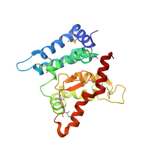
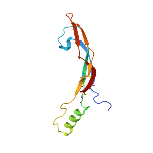
The Structure of the Glial Cell Line-Derived Neurotrophic Factor-Coreceptor Complex: Insights Into Ret Signaling and Heparin Binding.

Parkash, V.Leppanen, V.-M.Virtanen, H.Jurvansuu, J.-M.Bespalov, M.M.Sidorova, Y.A.Runeberg-Roos, P.Saarma, M.Goldman, A.

(2008) J Biol Chem 283: 35164

  • DOI: https://doi.org/10.1074/jbc.M802543200
  • Primary Citation of Related Structures:  
    2V5E

  • PubMed Abstract: 

    Glial cell line-derived neurotrophic factor (GDNF), a neuronal survival factor, binds its co-receptor GDNF family receptor alpha1 (GFR alpha 1) in a 2:2 ratio and signals through the receptor tyrosine kinase RET. We have solved the GDNF(2).GFR alpha 1(2) complex structure at 2.35 A resolution in the presence of a heparin mimic, sucrose octasulfate. The structure of our GDNF(2).GFR alpha 1(2) complex and the previously published artemin(2).GFR alpha 3(2) complex are unlike in three ways. First, we have experimentally identified residues that differ in the ligand-GFR alpha interface between the two structures, in particular ones that buttress the key conserved Arg(GFR alpha)-Glu(ligand)-Arg(GFR alpha) interaction. Second, the flexible GDNF ligand "finger" loops fit differently into the GFR alphas, which are rigid. Third, and we believe most importantly, the quaternary structure of the two tetramers is dissimilar, because the angle between the two GDNF monomers is different. This suggests that the RET-RET interaction differs in different ligand(2)-co-receptor(2)-RET(2) heterohexamer complexes. Consistent with this, we showed that GDNF(2).GFR alpha1(2) and artemin(2).GFR alpha 3(2) signal differently in a mitogen-activated protein kinase assay. Furthermore, we have shown by mutagenesis and enzyme-linked immunosorbent assays of RET phosphorylation that RET probably interacts with GFR alpha 1 residues Arg-190, Lys-194, Arg-197, Gln-198, Lys-202, Arg-257, Arg-259, Glu-323, and Asp-324 upon both domains 2 and 3. Interestingly, in our structure, sucrose octasulfate also binds to the Arg(190)-Lys(202) region in GFR alpha 1 domain 2. This may explain how GDNF.GFR alpha 1 can mediate cell adhesion and how heparin might inhibit GDNF signaling through RET.


  • Organizational Affiliation

    Institute of Biotechnology, University of Helsinki, Helsinki, Finland.


Macromolecules
Find similar proteins by:  (by identity cutoff)  |  3D Structure
Entity ID: 1
MoleculeChains Sequence LengthOrganismDetailsImage
GDNF FAMILY RECEPTOR ALPHA-1200Rattus norvegicusMutation(s): 0 
UniProt
Find proteins for Q62997 (Rattus norvegicus)
Explore Q62997 
Go to UniProtKB:  Q62997
Entity Groups  
Sequence Clusters30% Identity50% Identity70% Identity90% Identity95% Identity100% Identity
UniProt GroupQ62997
Sequence Annotations
Expand
  • Reference Sequence
Find similar proteins by:  (by identity cutoff)  |  3D Structure
Entity ID: 2
MoleculeChains Sequence LengthOrganismDetailsImage
GLIAL CELL LINE-DERIVED NEUROTROPHIC FACTOR101Homo sapiensMutation(s): 0 
UniProt & NIH Common Fund Data Resources
Find proteins for P39905 (Homo sapiens)
Explore P39905 
Go to UniProtKB:  P39905
PHAROS:  P39905
GTEx:  ENSG00000168621 
Entity Groups  
Sequence Clusters30% Identity50% Identity70% Identity90% Identity95% Identity100% Identity
UniProt GroupP39905
Sequence Annotations
Expand
  • Reference Sequence
Oligosaccharides

Help

Entity ID: 3
MoleculeChains Length2D Diagram Glycosylation3D Interactions
1,3,4,6-tetra-O-sulfo-beta-D-fructofuranose-(2-1)-2,3,4,6-tetra-O-sulfonato-alpha-D-glucopyranose
C
2N/A
Glycosylation Resources
GlyTouCan:  G97555GV
GlyCosmos:  G97555GV
Biologically Interesting Molecules (External Reference) 1 Unique
Experimental Data & Validation

Experimental Data

  • Method: X-RAY DIFFRACTION
  • Resolution: 2.35 Å
  • R-Value Free: 0.237 
  • R-Value Work: 0.184 
  • R-Value Observed: 0.187 
  • Space Group: C 1 2 1
Unit Cell:
Length ( Å )Angle ( ˚ )
a = 58.9α = 90
b = 75.7β = 91.9
c = 105.5γ = 90
Software Package:
Software NamePurpose
REFMACrefinement
XSCALEdata scaling
PHASERphasing

Structure Validation

View Full Validation Report


Ligand Structure Quality Assessment 


Entry History 

Deposition Data

Revision History  (Full details and data files)

  • Version 1.0: 2008-10-21
    Type: Initial release
  • Version 1.1: 2014-02-05
    Changes: Derived calculations, Non-polymer description, Other, Refinement description, Source and taxonomy, Structure summary, Version format compliance
  • Version 2.0: 2020-07-29
    Type: Remediation
    Reason: Carbohydrate remediation
    Changes: Atomic model, Data collection, Derived calculations, Non-polymer description, Other, Structure summary
  • Version 2.1: 2023-12-13
    Changes: Data collection, Database references, Refinement description, Structure summary