Vitamin B12 Transport Proteins: Crystallographic Analysis of Beta-Axial Ligand Substitutions in Cobalamin Bound to Transcobalamin.
Wuerges, J., Geremia, S., Fedosov, S.N., Randaccio, L.(2007) IUBMB Life 59: 722
- PubMed: 17943552
- DOI: https://doi.org/10.1080/15216540701673413
- Primary Citation of Related Structures:
2V3N, 2V3P - PubMed Abstract:
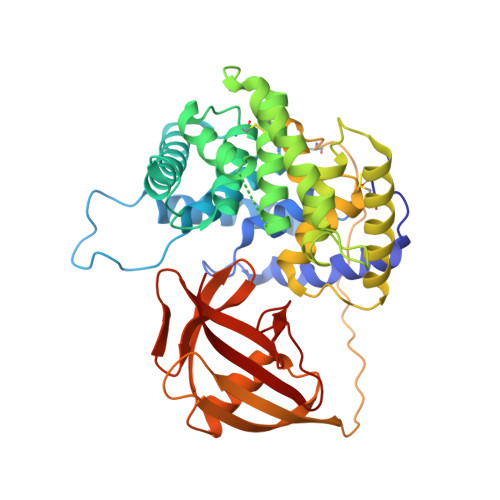
Cobalamin (Cbl, vitamin B12) is an essential micronutrient that is synthesized only by bacteria. Mammals have developed a complex system for internalization of this vitamin from the diet. Three binding proteins (haptocorrin, intrinsic factor, transcobalamin (TC)) and several specific cell surface receptors are involved in the process of intestinal absorption, plasma transport and cellular uptake. The recent literature on the binding proteins is briefly reviewed. A structural study is presented addressing a unique feature of TC among the three proteins, i.e., the displacement of the weak Co(III)-ligand H2O at the upper (or beta) axial side of H2O-Cbl by a histidine side chain. We have investigated crystallographically the beta-ligand exchange on Cbl bound to TC by crystallization of bovine holo-TC in the presence of either cyanide or sulfite. The resulting electron density maps show that the histidine side chain has been displaced by an exogenous ligand CN(-) or SO(3)(-2)to a lower extent than expected based on their higher affinity for Co and excess concentration with respect to histidine. This may reflect either reduced affinities of CN(-) and SO(3)(-2)or the advantageous binding of the protein-integrated His-residue when competing for the beta-site of Cbl bound to TC. The loop hosting the histidine residue appears more flexible after disruption of the coordination bond His-Cbl but no other differences are observed in the overall structure of holo-TC. These structural results are discussed in relation to a possible physiological role of histidine substitution for H2O and regarding the role of beta-conjugated Cbl-analogues recently proposed for targeted delivery of imaging agents.
Organizational Affiliation:
Centre of Excellence in Biocrystallography, Department of Chemical Sciences, University of Trieste, Trieste, Italy.