2SPT

DIFFERENCES IN THE METAL ION STRUCTURE BETWEEN SR-AND CA-PROTHROMBIN FRAGMENT 1


Experimental Data Snapshot

  • Method: X-RAY DIFFRACTION
  • Resolution: 2.50 Å
  • R-Value Observed: 0.167 

wwPDB Validation   3D Report Full Report


This is version 1.4 of the entry. See complete history


Literature

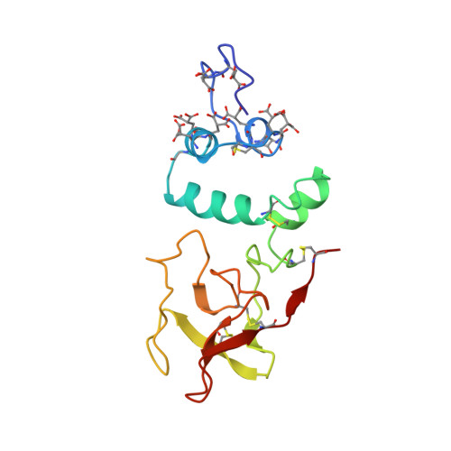
Differences in the metal ion structure between Sr- and Ca-prothrombin fragment 1.

Seshadri, T.P.Skrzypczak-Jankun, E.Yin, M.Tulinsky, A.

(1994) Biochemistry 33: 1087-1092

  • DOI: https://doi.org/10.1021/bi00171a006
  • Primary Citation of Related Structures:  
    2SPT

  • PubMed Abstract: 

    The structure of Sr-prothrombin fragment 1 has been solved and refined by restrained least-squares methods at 2.5-A resolution to a crystallographic R value of 0.167. The protein structure is very similar to that of Ca-fragment 1. A polymeric array of five Sr2+ ions separated by about 4.0 A is buried among six gamma-carboxyglutamic acid (Gla) residues; three other Sr2+ ions interact with other Gla residues and are located further apart. One of these was not found in the Ca-fragment 1 structure. The coordination of the Sr2+ ions resembles that of Ca2+, but there are some significant differences between them. The most notable is the lack of water coordination with Sr2+ ions and two conformations for Gla 8, which change the coordination of Sr-2 and Sr-3. A hexose moiety of an oligosaccharide was located in the vicinity of Asn101 that was flexibly disordered in Ca-fragment 1. The new Sr2+ ion found may be involved in metal ion phospholipid binding interactions along with Sr-1, and Sr-7, Sr-8.


  • Organizational Affiliation

    Department of Chemistry, Michigan State University, East Lansing 48824.


Macromolecules
Find similar proteins by:  (by identity cutoff)  |  3D Structure
Entity ID: 1
MoleculeChains Sequence LengthOrganismDetailsImage
PROTHROMBIN145Bos taurusMutation(s): 0 
EC: 3.4.21.5
UniProt
Find proteins for P00735 (Bos taurus)
Explore P00735 
Go to UniProtKB:  P00735
Entity Groups  
Sequence Clusters30% Identity50% Identity70% Identity90% Identity95% Identity100% Identity
UniProt GroupP00735
Sequence Annotations
Expand
  • Reference Sequence
Experimental Data & Validation

Experimental Data

  • Method: X-RAY DIFFRACTION
  • Resolution: 2.50 Å
  • R-Value Observed: 0.167 
  • Space Group: P 21 21 21
Unit Cell:
Length ( Å )Angle ( ˚ )
a = 39.69α = 90
b = 54.12β = 90
c = 131.17γ = 90
Software Package:
Software NamePurpose
PROFFTrefinement
PROLSQrefinement

Structure Validation

View Full Validation Report


Entry History 

Deposition Data

  • Released Date: 1994-05-31 
  • Deposition Author(s): Tulinsky, A.

Revision History  (Full details and data files)

  • Version 1.0: 1994-05-31
    Type: Initial release
  • Version 1.1: 2008-03-25
    Changes: Version format compliance
  • Version 1.2: 2011-07-13
    Changes: Non-polymer description, Version format compliance
  • Version 1.3: 2017-11-29
    Changes: Derived calculations, Other
  • Version 1.4: 2020-07-29
    Type: Remediation
    Reason: Carbohydrate remediation
    Changes: Data collection, Database references, Derived calculations, Structure summary