2RH7

Crystal Structures of the Luciferase and Green Fluorescent Protein from Renilla Reniformis


Experimental Data Snapshot

  • Method: X-RAY DIFFRACTION
  • Resolution: 1.50 Å
  • R-Value Free: 0.325 
  • R-Value Work: 0.291 

wwPDB Validation   3D Report Full Report


This is version 1.3 of the entry. See complete history


Literature

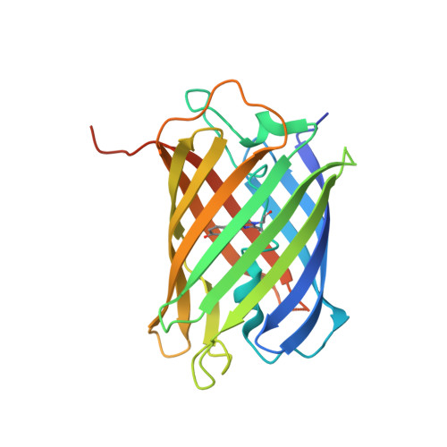
Crystal Structures of the Luciferase and Green Fluorescent Protein from Renilla reniformis.

Loening, A.M.Fenn, T.D.Gambhir, S.S.

(2007) J Mol Biol 374: 1017-1028

  • DOI: https://doi.org/10.1016/j.jmb.2007.09.078
  • Primary Citation of Related Structures:  
    2PSD, 2PSE, 2PSF, 2PSH, 2PSJ, 2RH7

  • PubMed Abstract: 

    Due to its ability to emit light, the luciferase from Renilla reniformis (RLuc) is widely employed in molecular biology as a reporter gene in cell culture experiments and small animal imaging. To accomplish this bioluminescence, the 37-kDa enzyme catalyzes the degradation of its substrate coelenterazine in the presence of molecular oxygen, resulting in the product coelenteramide, carbon dioxide, and the desired photon of light. We successfully crystallized a stabilized variant of this important protein (RLuc8) and herein present the first structures for any coelenterazine-using luciferase. These structures are based on high-resolution data measured to 1.4 A and demonstrate a classic alpha/beta-hydrolase fold. We also present data of a coelenteramide-bound luciferase and reason that this structure represents a secondary conformational form following shift of the product out of the primary active site. During the course of this work, the structure of the luciferase's accessory green fluorescent protein (RrGFP) was also determined and shown to be highly similar to that of Aequorea victoria GFP.


  • Organizational Affiliation

    Molecular Imaging Program at Stanford, Department of Radiology and Bio-X Program, The James H. Clark Center, Stanford University School of Medicine, 318 Campus Drive, Clark E150, Stanford, CA 94305-5427, USA.


Macromolecules
Find similar proteins by:  (by identity cutoff)  |  3D Structure
Entity ID: 1
MoleculeChains Sequence LengthOrganismDetailsImage
Green fluorescent protein
A, B, C, D
239Renilla reniformisMutation(s): 1 
Gene Names: GFP
UniProt
Find proteins for Q963I9 (Renilla reniformis)
Explore Q963I9 
Go to UniProtKB:  Q963I9
Entity Groups  
Sequence Clusters30% Identity50% Identity70% Identity90% Identity95% Identity100% Identity
UniProt GroupQ963I9
Sequence Annotations
Expand
  • Reference Sequence
Small Molecules
Modified Residues  1 Unique
IDChains TypeFormula2D DiagramParent
CRO
Query on CRO
A, B, C, D
L-PEPTIDE LINKINGC15 H17 N3 O5THR, TYR, GLY
Experimental Data & Validation

Experimental Data

  • Method: X-RAY DIFFRACTION
  • Resolution: 1.50 Å
  • R-Value Free: 0.325 
  • R-Value Work: 0.291 
  • Space Group: P 21 21 2
Unit Cell:
Length ( Å )Angle ( ˚ )
a = 73.744α = 90
b = 85.405β = 90
c = 158.425γ = 90
Software Package:
Software NamePurpose
PHASERphasing
REFMACrefinement
DENZOdata reduction
SCALEPACKdata scaling

Structure Validation

View Full Validation Report


Entry History 

Deposition Data

Revision History  (Full details and data files)

  • Version 1.0: 2007-10-23
    Type: Initial release
  • Version 1.1: 2011-07-13
    Changes: Version format compliance
  • Version 1.2: 2023-08-30
    Changes: Advisory, Data collection, Database references, Refinement description
  • Version 1.3: 2023-11-15
    Changes: Data collection