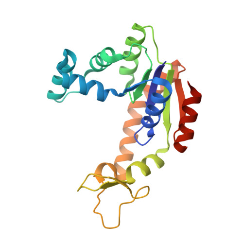
Intrinsic motions along an enzymatic reaction trajectory.
Henzler-Wildman, K.A., Thai, V., Lei, M., Ott, M., Wolf-Watz, M., Fenn, T., Pozharski, E., Wilson, M.A., Petsko, G.A., Karplus, M., Hubner, C.G., Kern, D.(2007) Nature 450: 838-844
- PubMed: 18026086
- DOI: https://doi.org/10.1038/nature06410
- Primary Citation of Related Structures:
2RGX, 2RH5 - PubMed Abstract:
The mechanisms by which enzymes achieve extraordinary rate acceleration and specificity have long been of key interest in biochemistry. It is generally recognized that substrate binding coupled to conformational changes of the substrate-enzyme complex aligns the reactive groups in an optimal environment for efficient chemistry. Although chemical mechanisms have been elucidated for many enzymes, the question of how enzymes achieve the catalytically competent state has only recently become approachable by experiment and computation. Here we show crystallographic evidence for conformational substates along the trajectory towards the catalytically competent 'closed' state in the ligand-free form of the enzyme adenylate kinase. Molecular dynamics simulations indicate that these partially closed conformations are sampled in nanoseconds, whereas nuclear magnetic resonance and single-molecule fluorescence resonance energy transfer reveal rare sampling of a fully closed conformation occurring on the microsecond-to-millisecond timescale. Thus, the larger-scale motions in substrate-free adenylate kinase are not random, but preferentially follow the pathways that create the configuration capable of proficient chemistry. Such preferred directionality, encoded in the fold, may contribute to catalysis in many enzymes.
Organizational Affiliation:
Department of Biochemistry and Howard Hughes Medical Institute, Brandeis University, Waltham, Massachusetts 02454, USA.