Structures of rat cytosolic PEPCK: insight into the mechanism of phosphorylation and decarboxylation of oxaloacetic acid.
Sullivan, S.M., Holyoak, T.(2007) Biochemistry 46: 10078-10088
- PubMed: 17685635
- DOI: https://doi.org/10.1021/bi701038x
- Primary Citation of Related Structures:
2QEW, 2QEY, 2QF1, 2QF2 - PubMed Abstract:
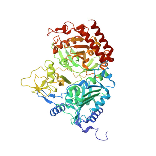
The structures of the rat cytosolic isoform of phosphoenolpyruvate carboxykinase (PEPCK) reported in the PEPCK-Mn2+, -Mn2+-oxaloacetic acid (OAA), -Mn2+-OAA-Mn2+-guanosine-5'-diphosphate (GDP), and -Mn2+-Mn2+-guanosine-5'-tri-phosphate (GTP) complexes provide insight into the mechanism of phosphoryl transfer and decarboxylation mediated by this enzyme. OAA is observed to bind in a number of different orientations coordinating directly to the active site metal. The Mn2+-OAA and Mn2+-OAA-Mn2+GDP structures illustrate inner-sphere coordination of OAA to the manganese ion through the displacement of two of the three water molecules coordinated to the metal in the holo-enzyme by the C3 and C4 carbonyl oxygens. In the PEPCK-Mn2+-OAA complex, an alternate bound conformation of OAA is present. In this conformation, in addition to the previous interactions, the C1 carboxylate is directly coordinated to the active site Mn2+, displacing all of the waters coordinated to the metal in the holo-enzyme. In the PEPCK-Mn2+-GTP structure, the same water molecule displaced by the C1 carboxylate of OAA is displaced by one of the gamma-phosphate oxygens of the triphosphate nucleotide. The structures are consistent with a mechanism of direct in-line phosphoryl transfer, supported by the observed stereochemistry of the reaction. In the catalytically competent binding mode, the C1 carboxylate of OAA is sandwiched between R87 and R405 in an environment that would serve to facilitate decarboxylation. In the reverse reaction, these two arginines would form the CO2 binding site. Comparison of the Mn2+-OAA-Mn2+GDP and Mn2+-Mn2+GTP structures illustrates a marked difference in the bound conformations of the nucleotide substrates in which the GTP nucleotide is bound in a high-energy state resulting from the eclipsing of all three of the phosphoryl groups along the triphosphate chain. This contrasts a previously determined structure of PEPCK in complex with a triphosphate nucleotide analogue in which the analogue mirrors the conformation of GDP as opposed to GTP. Last, the structures illustrate a correlation between conformational changes in the P-loop, the nucleotide binding site, and the active site lid that are important for catalysis.
Organizational Affiliation:
Department of Biochemistry and Molecular Biology, The University of Kansas Medical Center, Kansas City, Kansas 66160, USA.