2QES

Crystal structure of the ribosome inactivating protein PDL4 from P. dioica leaves in complex with adenine


Experimental Data Snapshot

  • Method: X-RAY DIFFRACTION
  • Resolution: 1.24 Å
  • R-Value Free: 0.145 
  • R-Value Work: 0.116 

wwPDB Validation   3D Report Full Report


This is version 1.2 of the entry. See complete history


Literature

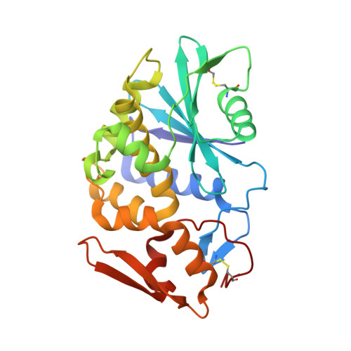
Atomic resolution (1.1 A) structure of the ribosome-inactivating protein PD-L4 from Phytolacca dioica L. leaves

Ruggiero, A.Chambery, A.Di Maro, A.Parente, A.Berisio, R.

(2008) Proteins 71: 8-15

  • DOI: https://doi.org/10.1002/prot.21712
  • Primary Citation of Related Structures:  
    2QES, 2QET, 2Z4U, 2Z53

  • PubMed Abstract: 

    The ribosome inactivating protein PD-L4 from Phytolacca dioica is a N-beta-glycosidase, probably involved in plant defence. The crystal structures of wild type PD-L4 and of the S211A PD-L4 mutant with significantly decreased catalytic activity were determined at atomic resolution. To determine the structural determinants for the reduced activity of S211A PD-L4, both forms have also been co-crystallized with adenine, the major product of PD-L4 catalytic reaction. In the structure of the S211A mutant, the cavity formed by the lack of the Ser hydroxyl group is filled by a water molecule; the insertion of this non-isosteric group leads to small albeit concerted changes in the tightly packed active site of the enzyme. These changes have been correlated to the different activity of the mutant enzyme. This work highlights the importance of atomic resolution studies for the deep understanding of enzymatic properties.


  • Organizational Affiliation

    Istituto di Biostrutture e Bioimmagini, C.N.R., Napoli I-80134, Italy.


Macromolecules
Find similar proteins by:  (by identity cutoff)  |  3D Structure
Entity ID: 1
MoleculeChains Sequence LengthOrganismDetailsImage
Ribosome-inactivating protein PD-L4261Phytolacca dioicaMutation(s): 0 
EC: 3.2.2.22
UniProt
Find proteins for P84854 (Phytolacca dioica)
Explore P84854 
Go to UniProtKB:  P84854
Entity Groups  
Sequence Clusters30% Identity50% Identity70% Identity90% Identity95% Identity100% Identity
UniProt GroupP84854
Sequence Annotations
Expand
  • Reference Sequence
Small Molecules
Ligands 1 Unique
IDChains Name / Formula / InChI Key2D Diagram3D Interactions
ADE
Query on ADE

Download Ideal Coordinates CCD File 
B [auth A]ADENINE
C5 H5 N5
GFFGJBXGBJISGV-UHFFFAOYSA-N
Experimental Data & Validation

Experimental Data

  • Method: X-RAY DIFFRACTION
  • Resolution: 1.24 Å
  • R-Value Free: 0.145 
  • R-Value Work: 0.116 
  • Space Group: P 21 21 21
Unit Cell:
Length ( Å )Angle ( ˚ )
a = 43.521α = 90
b = 58.813β = 90
c = 98.774γ = 90
Software Package:
Software NamePurpose
HKL-2000data collection
IlMilionemodel building
SHELXL-97refinement
HKL-2000data reduction
HKL-2000data scaling
IlMilionephasing

Structure Validation

View Full Validation Report


Entry History 

Deposition Data

Revision History  (Full details and data files)

  • Version 1.0: 2008-02-26
    Type: Initial release
  • Version 1.1: 2011-07-13
    Changes: Version format compliance
  • Version 1.2: 2023-10-25
    Changes: Data collection, Database references, Derived calculations, Refinement description