2QBQ

Crystal structure of ptp1b-inhibitor complex


Experimental Data Snapshot

  • Method: X-RAY DIFFRACTION
  • Resolution: 2.10 Å
  • R-Value Free: 0.226 
  • R-Value Work: 0.194 
  • R-Value Observed: 0.194 

wwPDB Validation   3D Report Full Report


Ligand Structure Quality Assessment 


This is version 1.2 of the entry. See complete history


Literature

Structure-based optimization of protein tyrosine phosphatase 1B inhibitors: from the active site to the second phosphotyrosine binding site.

Wilson, D.P.Wan, Z.K.Xu, W.X.Kirincich, S.J.Follows, B.C.Joseph-McCarthy, D.Foreman, K.Moretto, A.Wu, J.Zhu, M.Binnun, E.Zhang, Y.L.Tam, M.Erbe, D.V.Tobin, J.Xu, X.Leung, L.Shilling, A.Tam, S.Y.Mansour, T.S.Lee, J.

(2007) J Med Chem 50: 4681-4698

  • DOI: https://doi.org/10.1021/jm0702478
  • Primary Citation of Related Structures:  
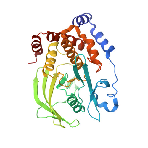
    2QBP, 2QBQ, 2QBR, 2QBS

  • PubMed Abstract: 

    Protein tyrosine phosphatase 1B (PTP1B) is a negative regulator of the insulin and leptin receptor pathways and thus an attractive therapeutic target for diabetes and obesity. Starting with a high micromolar lead compound, structure-based optimization of novel PTP1B inhibitors by extension of the molecule from the enzyme active site into the second phosphotyrosine binding site is described. Medicinal chemistry, guided by X-ray complex structure and molecular modeling, has yielded low nanomolar PTP1B inhibitors in an efficient manner. Compounds from this chemical series were found to be actively transported into hepatocytes. This active uptake into target tissues could be one of the possible avenues to overcome the poor membrane permeability of PTP1B inhibitors.


  • Organizational Affiliation

    Chemical and Screening Sciences, and Cardiovascular and Metabolic Diseases, Wyeth Research, 200 Cambridge Park Drive, Cambridge, Massachusetts 02140, USA.


Macromolecules
Find similar proteins by:  (by identity cutoff)  |  3D Structure
Entity ID: 1
MoleculeChains Sequence LengthOrganismDetailsImage
Tyrosine-protein phosphatase non-receptor type 1299Homo sapiensMutation(s): 0 
Gene Names: PTPN1PTP1B
EC: 3.1.3.48
UniProt & NIH Common Fund Data Resources
Find proteins for P18031 (Homo sapiens)
Explore P18031 
Go to UniProtKB:  P18031
PHAROS:  P18031
GTEx:  ENSG00000196396 
Entity Groups  
Sequence Clusters30% Identity50% Identity70% Identity90% Identity95% Identity100% Identity
UniProt GroupP18031
Sequence Annotations
Expand
  • Reference Sequence
Small Molecules
Ligands 1 Unique
IDChains Name / Formula / InChI Key2D Diagram3D Interactions
4B3
Query on 4B3

Download Ideal Coordinates CCD File 
B [auth A]4-BROMO-3-(CARBOXYMETHOXY)-5-{3-[(3,3,5,5-TETRAMETHYLCYCLOHEXYL)AMINO]PHENYL}THIOPHENE-2-CARBOXYLIC ACID
C23 H28 Br N O5 S
OSQARHVGUZHHGT-UHFFFAOYSA-N
Binding Affinity Annotations 
IDSourceBinding Affinity
4B3 BindingDB:  2QBQ Ki: min: 26, max: 36 (nM) from 2 assay(s)
PDBBind:  2QBQ Ki: 36 (nM) from 1 assay(s)
Binding MOAD:  2QBQ Ki: 36 (nM) from 1 assay(s)
Experimental Data & Validation

Experimental Data

  • Method: X-RAY DIFFRACTION
  • Resolution: 2.10 Å
  • R-Value Free: 0.226 
  • R-Value Work: 0.194 
  • R-Value Observed: 0.194 
  • Space Group: P 31 2 1
Unit Cell:
Length ( Å )Angle ( ˚ )
a = 88.345α = 90
b = 88.345β = 90
c = 104.21γ = 120
Software Package:
Software NamePurpose
CNSrefinement
CNSphasing

Structure Validation

View Full Validation Report


Ligand Structure Quality Assessment 


Entry History 

Deposition Data

  • Released Date: 2008-03-18 
  • Deposition Author(s): Xu, W.

Revision History  (Full details and data files)

  • Version 1.0: 2008-03-18
    Type: Initial release
  • Version 1.1: 2011-07-13
    Changes: Version format compliance
  • Version 1.2: 2024-02-21
    Changes: Data collection, Database references, Derived calculations