2PRM

The structures of apo- and inhibitor bound human dihydroorotate dehydrogenase reveal conformational flexibility within the inhibitor binding site


Experimental Data Snapshot

  • Method: X-RAY DIFFRACTION
  • Resolution: 3.00 Å
  • R-Value Free: 0.253 
  • R-Value Work: 0.190 
  • R-Value Observed: 0.193 

wwPDB Validation   3D Report Full Report


Ligand Structure Quality Assessment 


This is version 1.3 of the entry. See complete history


Literature

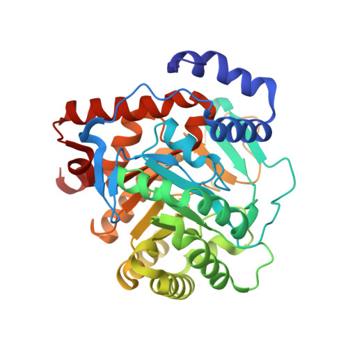
The structures of human dihydroorotate dehydrogenase with and without inhibitor reveal conformational flexibility in the inhibitor and substrate binding sites

Walse, B.Dufe, V.T.Svensson, B.Fritzson, I.Dahlberg, L.Khairoullina, A.Wellmar, U.Al-Karadaghi, S.

(2008) Biochemistry 47: 8929-8936

  • DOI: https://doi.org/10.1021/bi8003318
  • Primary Citation of Related Structures:  
    2PRH, 2PRL, 2PRM

  • PubMed Abstract: 

    Inhibitors of dihydroorotate dehydrogenase (DHODH) have been suggested for the treatment of rheumatoid arthritis, psoriasis, autoimmune diseases, Plasmodium, and bacterial and fungal infections. Here we present the structures of N-terminally truncated (residues Met30-Arg396) DHODH in complex with two inhibitors: a brequinar analogue (6) and a novel inhibitor (a fenamic acid derivative) (7), as well as the first structure of the enzyme to be characterized without any bound inhibitor. It is shown that 7 uses the "standard" brequinar binding mode and, in addition, interacts with Tyr356, a residue conserved in most class 2 DHODH proteins. Compared to the inhibitor-free structure, some of the amino acid side chains in the tunnel in which brequinar binds and which was suggested to be the binding site of ubiquinone undergo changes in conformation upon inhibitor binding. Using our data, the loop regions of residues Leu68-Arg72 and Asn212-Leu224, which were disordered in previously studied human DHODH structures, could be built into the electron density. The first of these loops, which is located at the entrance to the inhibitor-binding pocket, shows different conformations in the three structures, suggesting that it may interfere with inhibitor/cofactor binding. The second loop has been suggested to control the access of dihydroorotate to the active site of the enzyme and may be an important player in the enzymatic reaction. These observations provide new insights into the dynamic features of the DHODH reaction and suggest new approaches to the design of inhibitors against DHODH.


  • Organizational Affiliation

    SARomics AB, P.O. Box 724, SE-220 07 Lund, Sweden. bjorn.walse@saromics.com


Macromolecules
Find similar proteins by:  (by identity cutoff)  |  3D Structure
Entity ID: 1
MoleculeChains Sequence LengthOrganismDetailsImage
Dihydroorotate dehydrogenase, mitochondrial367Homo sapiensMutation(s): 0 
Gene Names: DHODH
EC: 1.3.99.11
Membrane Entity: Yes 
UniProt & NIH Common Fund Data Resources
Find proteins for Q02127 (Homo sapiens)
Explore Q02127 
Go to UniProtKB:  Q02127
PHAROS:  Q02127
GTEx:  ENSG00000102967 
Entity Groups  
Sequence Clusters30% Identity50% Identity70% Identity90% Identity95% Identity100% Identity
UniProt GroupQ02127
Sequence Annotations
Expand
  • Reference Sequence
Small Molecules
Ligands 2 Unique
IDChains Name / Formula / InChI Key2D Diagram3D Interactions
FMN
Query on FMN

Download Ideal Coordinates CCD File 
B [auth A]FLAVIN MONONUCLEOTIDE
C17 H21 N4 O9 P
FVTCRASFADXXNN-SCRDCRAPSA-N
ORO
Query on ORO

Download Ideal Coordinates CCD File 
C [auth A]OROTIC ACID
C5 H4 N2 O4
PXQPEWDEAKTCGB-UHFFFAOYSA-N
Experimental Data & Validation

Experimental Data

  • Method: X-RAY DIFFRACTION
  • Resolution: 3.00 Å
  • R-Value Free: 0.253 
  • R-Value Work: 0.190 
  • R-Value Observed: 0.193 
  • Space Group: P 32 2 1
Unit Cell:
Length ( Å )Angle ( ˚ )
a = 90.05α = 90
b = 90.05β = 90
c = 123.2γ = 120
Software Package:
Software NamePurpose
XSCALEdata scaling
REFMACrefinement
PDB_EXTRACTdata extraction
MAR345dtbdata collection
XDSdata reduction
CNSphasing

Structure Validation

View Full Validation Report


Ligand Structure Quality Assessment 


Entry History 

Deposition Data

Revision History  (Full details and data files)

  • Version 1.0: 2008-05-20
    Type: Initial release
  • Version 1.1: 2011-07-13
    Changes: Version format compliance
  • Version 1.2: 2018-03-07
    Changes: Data collection
  • Version 1.3: 2023-08-30
    Changes: Data collection, Database references, Derived calculations, Refinement description