


Crystal Structure Analysis of the West Nile virus envelope (E) protein domain III
Liu, J., Gao, F., Yuan, F., Gao, G.F.To be published.
Experimental Data Snapshot
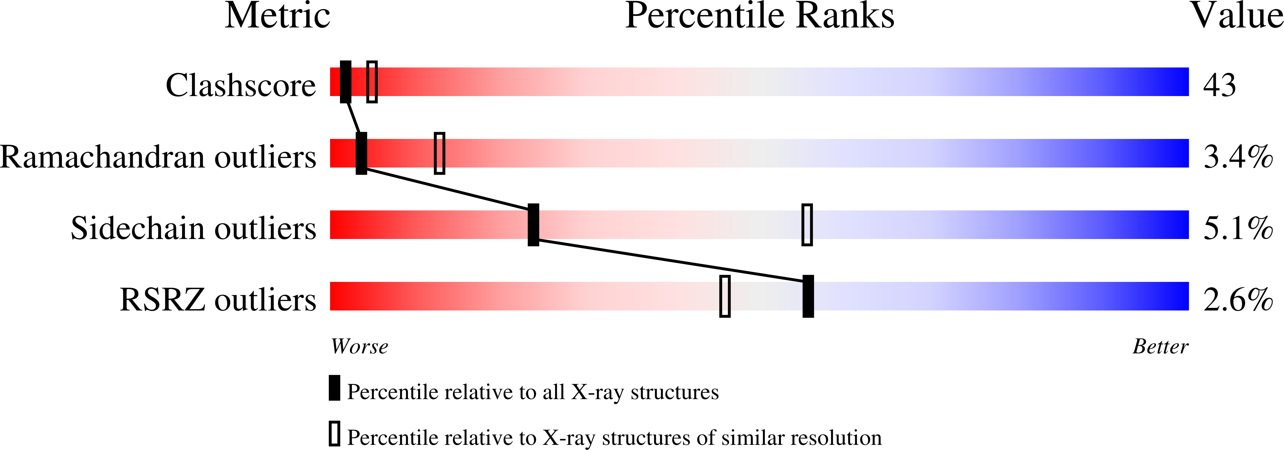
wwPDB Validation 3D Report Full Report
Entity ID: 1 | |||||
|---|---|---|---|---|---|
| Molecule | Chains | Sequence Length | Organism | Details | Image |
| Genome polyprotein | 117 | West Nile virus | Mutation(s): 0 | 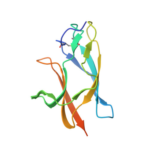 | |
UniProt | |||||
Find proteins for P06935 (West Nile virus) Explore P06935 Go to UniProtKB: P06935 | |||||
Entity Groups | |||||
| Sequence Clusters | 30% Identity50% Identity70% Identity90% Identity95% Identity100% Identity | ||||
| UniProt Group | P06935 | ||||
Sequence AnnotationsExpand | |||||
| |||||
| Length ( Å ) | Angle ( ˚ ) |
|---|---|
| a = 52.56 | α = 90 |
| b = 59.69 | β = 90 |
| c = 95.03 | γ = 90 |
| Software Name | Purpose |
|---|---|
| CrystalClear | data collection |
| CNS | refinement |
| DENZO | data reduction |
| SCALEPACK | data scaling |
| CNS | phasing |
RCSB PDB (citation) is hosted by
RCSB PDB is a member of the

