2P2D

Crystal Structure and Allosteric Regulation of the Cytoplasmic Escherichia coli L-Asparaginase I


Experimental Data Snapshot

  • Method: X-RAY DIFFRACTION
  • Resolution: 1.89 Å
  • R-Value Free: 0.207 
  • R-Value Work: 0.184 

wwPDB Validation   3D Report Full Report


This is version 1.3 of the entry. See complete history


Literature

Crystal Structure and Allosteric Regulation of the Cytoplasmic Escherichia colil-Asparaginase I

Yun, M.-K.Nourse, A.White, S.W.Rock, C.O.Heath, R.J.

(2007) J Mol Biol 369: 794-811

  • DOI: https://doi.org/10.1016/j.jmb.2007.03.061
  • Primary Citation of Related Structures:  
    2HIM, 2P2D, 2P2N

  • PubMed Abstract: 

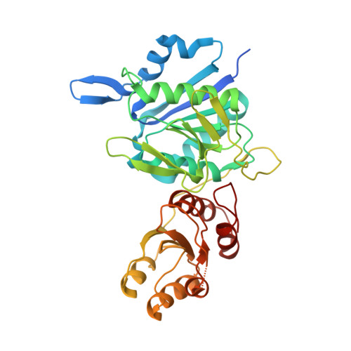
    AnsA is the cytoplasmic asparaginase from Escherichia coli involved in intracellular asparagine utilization. Analytical ultracentifugation and X-ray crystallography reveal that AnsA forms a tetrameric structure as a dimer of two intimate dimers. Kinetic analysis of the enzyme reveals that AnsA is positively cooperative, displaying a sigmoidal substrate dependence curve with an [S](0.5) of 1 mM L-asparagine and a Hill coefficient (n(H)) of 2.6. Binding of L-asparagine to an allosteric site was observed in the crystal structure concomitant with a reorganization of the quarternary structure, relative to the apo enzyme. The carboxyl group of the bound asparagine makes salt bridges and hydrogen bonds to Arg240, while the N(delta2) nitrogen interacts with Thr162. Mutation of Arg240 to Ala increases the [S](0.5) value to 5.9 mM, presumably by reducing the affinity of the site for L-asparagine, although the enzyme retains cooperativity. Mutation of Thr162 to Ala results in an active enzyme with no cooperativity. Transmission of the signal from the allosteric site to the active site appears to involve subtle interactions at the dimer-dimer interface and relocation of Gln118 into the vicinity of the active site to position the probable catalytic water molecule. These data define the structural basis for the cooperative regulation of the intracellular asparaginase that is required for proper functioning within the cell.


  • Organizational Affiliation

    Department of Structural Biology, St Jude Children's Research Hospital, Memphis, TN 38105, USA.


Macromolecules
Find similar proteins by:  (by identity cutoff)  |  3D Structure
Entity ID: 1
MoleculeChains Sequence LengthOrganismDetailsImage
L-ASPARAGINASE I
A, B, C, D
358Escherichia coliMutation(s): 0 
Gene Names: ansA
EC: 3.5.1.1
UniProt
Find proteins for P0A962 (Escherichia coli (strain K12))
Explore P0A962 
Go to UniProtKB:  P0A962
Entity Groups  
Sequence Clusters30% Identity50% Identity70% Identity90% Identity95% Identity100% Identity
UniProt GroupP0A962
Sequence Annotations
Expand
  • Reference Sequence
Small Molecules
Ligands 1 Unique
IDChains Name / Formula / InChI Key2D Diagram3D Interactions
GOL
Query on GOL

Download Ideal Coordinates CCD File 
E [auth A]
F [auth A]
G [auth A]
H [auth B]
I [auth B]
E [auth A],
F [auth A],
G [auth A],
H [auth B],
I [auth B],
J [auth B],
K [auth C],
L [auth C],
M [auth C],
N [auth D],
O [auth D],
P [auth D]
GLYCEROL
C3 H8 O3
PEDCQBHIVMGVHV-UHFFFAOYSA-N
Experimental Data & Validation

Experimental Data

  • Method: X-RAY DIFFRACTION
  • Resolution: 1.89 Å
  • R-Value Free: 0.207 
  • R-Value Work: 0.184 
  • Space Group: P 1 21 1
Unit Cell:
Length ( Å )Angle ( ˚ )
a = 57.763α = 90
b = 151.586β = 88.63
c = 87.142γ = 90
Software Package:
Software NamePurpose
MOLREPphasing
CNSrefinement
HKL-2000data reduction
HKL-2000data scaling

Structure Validation

View Full Validation Report


Entry History 

Deposition Data

Revision History  (Full details and data files)

  • Version 1.0: 2007-05-15
    Type: Initial release
  • Version 1.1: 2008-05-01
    Changes: Version format compliance
  • Version 1.2: 2011-07-13
    Changes: Non-polymer description, Version format compliance
  • Version 1.3: 2023-08-30
    Changes: Data collection, Database references, Derived calculations, Refinement description