


Crystal structure of phosphoribosyltransferase from Corynebacterium diphtheriae
Chang, C., Li, H., Clancy, S., Joachimiak, A.To be published.
Experimental Data Snapshot
wwPDB Validation 3D Report Full Report

Entity ID: 1 | |||||
|---|---|---|---|---|---|
| Molecule | Chains | Sequence Length | Organism | Details | Image |
| Phosphoribosyltransferase | 180 | Corynebacterium diphtheriae NCTC 13129 | Mutation(s): 3 Gene Names: pyrE, DIP2097 EC: 2.4.2.10 | 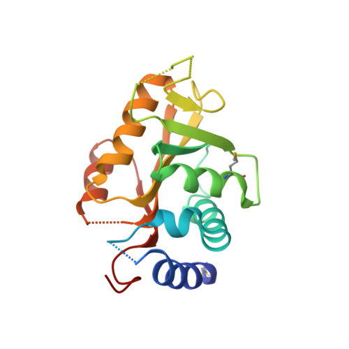 | |
UniProt | |||||
Find proteins for Q6NF12 (Corynebacterium diphtheriae (strain ATCC 700971 / NCTC 13129 / Biotype gravis)) Explore Q6NF12 Go to UniProtKB: Q6NF12 | |||||
Entity Groups | |||||
| Sequence Clusters | 30% Identity50% Identity70% Identity90% Identity95% Identity100% Identity | ||||
| UniProt Group | Q6NF12 | ||||
Sequence AnnotationsExpand | |||||
| |||||
| Modified Residues 1 Unique | |||||
|---|---|---|---|---|---|
| ID | Chains | Type | Formula | 2D Diagram | Parent |
| MSE Query on MSE | A, B | L-PEPTIDE LINKING | C5 H11 N O2 Se |  | MET |
| Length ( Å ) | Angle ( ˚ ) |
|---|---|
| a = 72.438 | α = 90 |
| b = 103.709 | β = 90 |
| c = 39.417 | γ = 90 |
| Software Name | Purpose |
|---|---|
| REFMAC | refinement |
| SBC-Collect | data collection |
| HKL-3000 | data reduction |
| HKL-3000 | data scaling |
| HKL-3000 | phasing |
RCSB PDB (citation) is hosted by
RCSB PDB is a member of the

