2OXB

Crystal structure of a cell-wall invertase (E203Q) from Arabidopsis thaliana in complex with sucrose


Experimental Data Snapshot

  • Method: X-RAY DIFFRACTION
  • Resolution: 2.60 Å
  • R-Value Free: 0.268 
  • R-Value Work: 0.205 
  • R-Value Observed: 0.205 

wwPDB Validation   3D Report Full Report


Ligand Structure Quality Assessment 


This is version 2.2 of the entry. See complete history


Literature

An alternate sucrose binding mode in the E203Q Arabidopsis invertase mutant: An X-ray crystallography and docking study.

Matrai, J.Lammens, W.Jonckheer, A.Le Roy, K.Rabijns, A.Van den Ende, W.De Maeyer, M.

(2007) Proteins 71: 552-564

  • DOI: https://doi.org/10.1002/prot.21700
  • Primary Citation of Related Structures:  
    2OXB

  • PubMed Abstract: 

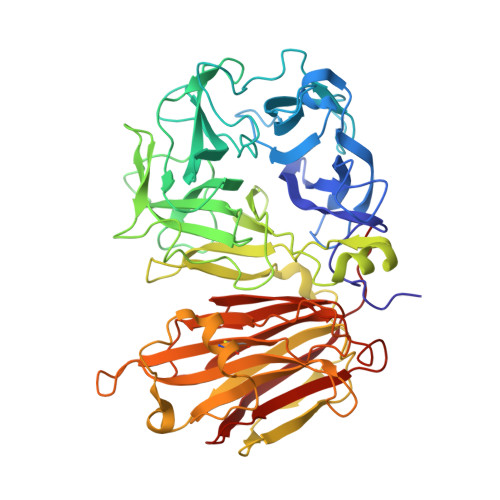
    In the present study, we report on the X-ray crystallographic structure of a GH32 invertase mutant, (i.e., the Arabidopsis thaliana cell-wall invertase 1-E203Q, AtcwINV1-mutant) in complex with sucrose. This structure was solved to reveal the features of sugar binding in the catalytic pocket. However, as demonstrated by the X-ray structure the sugar binding and the catalytic pocket arrangement is significantly altered as compared with what was expected based on previous X-ray structures on GH-J clan enzymes. We performed a series of docking and molecular dynamics simulations on various derivatives of AtcwINV1 to reveal the reasons behind this modified sugar binding. Our results demonstrate that the E203Q mutation introduced into the catalytic pocket triggers conformational changes that alter the wild type substrate binding. In addition, this study also reveals the putative productive sucrose binding modus in the wild type enzyme.


  • Organizational Affiliation

    Laboratory of Biomolecular Modelling and BioMacS, Department of Chemistry, Katholieke Universiteit Leuven, Belgium.


Macromolecules
Find similar proteins by:  (by identity cutoff)  |  3D Structure
Entity ID: 1
MoleculeChains Sequence LengthOrganismDetailsImage
Beta-fructofuranosidase537Arabidopsis thalianaMutation(s): 1 
Gene Names: ATBFRUCT1
EC: 3.2.1.26
UniProt
Find proteins for Q43866 (Arabidopsis thaliana)
Explore Q43866 
Go to UniProtKB:  Q43866
Entity Groups  
Sequence Clusters30% Identity50% Identity70% Identity90% Identity95% Identity100% Identity
UniProt GroupQ43866
Sequence Annotations
Expand
  • Reference Sequence
Oligosaccharides

Help

Entity ID: 2
MoleculeChains Length2D Diagram Glycosylation3D Interactions
beta-D-fructofuranose-(2-1)-alpha-D-glucopyranose
B
2N/A
Glycosylation Resources
GlyTouCan:  G05551OP
GlyCosmos:  G05551OP
Biologically Interesting Molecules (External Reference) 1 Unique
Experimental Data & Validation

Experimental Data

  • Method: X-RAY DIFFRACTION
  • Resolution: 2.60 Å
  • R-Value Free: 0.268 
  • R-Value Work: 0.205 
  • R-Value Observed: 0.205 
  • Space Group: P 32
Unit Cell:
Length ( Å )Angle ( ˚ )
a = 106.454α = 90
b = 106.454β = 90
c = 50.548γ = 120
Software Package:
Software NamePurpose
CNSrefinement
MAR345dtbdata collection
DENZOdata reduction
SCALEPACKdata scaling
CNSphasing

Structure Validation

View Full Validation Report


Ligand Structure Quality Assessment 


Entry History 

Deposition Data

Revision History  (Full details and data files)

  • Version 1.0: 2008-01-22
    Type: Initial release
  • Version 1.1: 2011-07-13
    Changes: Non-polymer description, Version format compliance
  • Version 2.0: 2020-07-29
    Type: Remediation
    Reason: Carbohydrate remediation
    Changes: Atomic model, Data collection, Derived calculations, Non-polymer description, Structure summary
  • Version 2.1: 2021-10-20
    Changes: Database references, Structure summary
  • Version 2.2: 2023-08-30
    Changes: Data collection, Refinement description