


X-ray structure of peptide-binding domain of Heat shock 70 kDa protein D precursor from C.elegans
Osipiuk, J., Duggan, E., Gu, M., Voisine, C., Morimoto, R.I., Joachimiak, A.To be published.
Experimental Data Snapshot
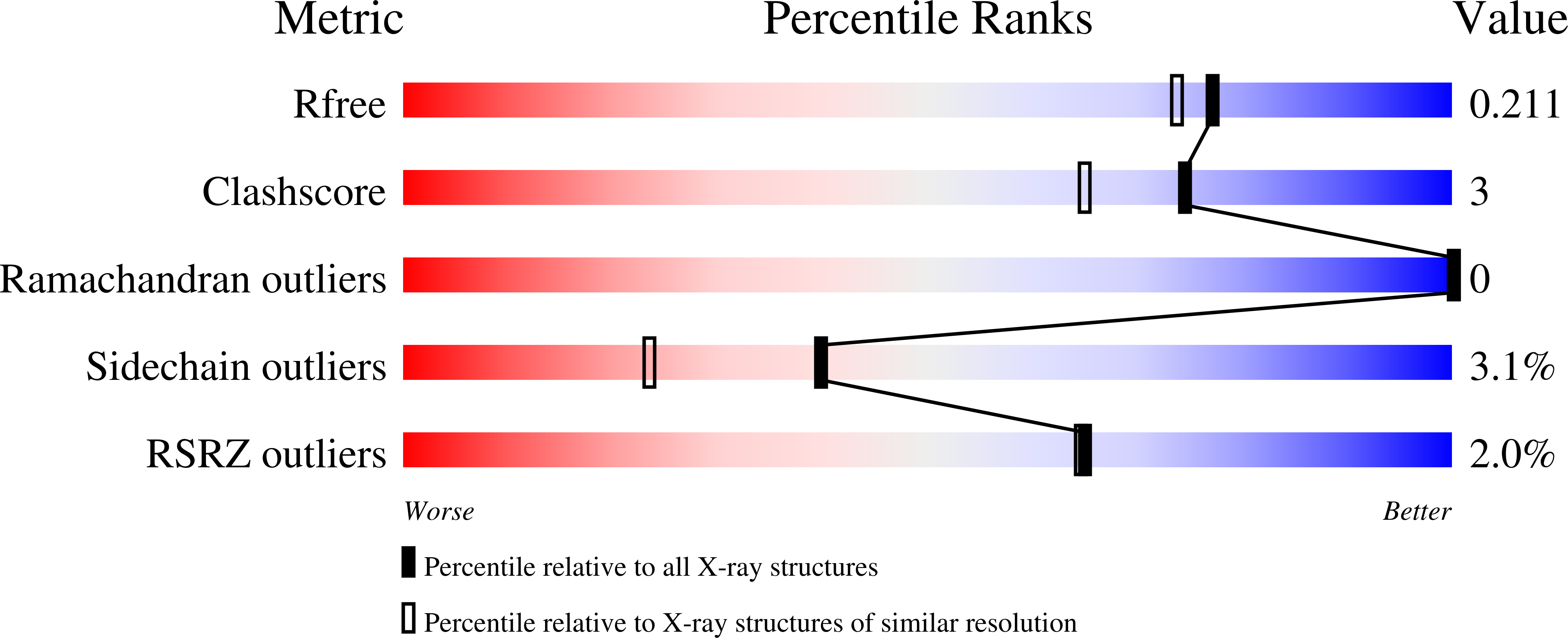
wwPDB Validation 3D Report Full Report
Entity ID: 1 | |||||
|---|---|---|---|---|---|
| Molecule | Chains | Sequence Length | Organism | Details | Image |
| Heat shock 70 kDa protein D | 152 | Caenorhabditis elegans | Mutation(s): 0 Gene Names: hsp-4, hsp70d |  | |
UniProt | |||||
Find proteins for P20163 (Caenorhabditis elegans) Explore P20163 Go to UniProtKB: P20163 | |||||
Entity Groups | |||||
| Sequence Clusters | 30% Identity50% Identity70% Identity90% Identity95% Identity100% Identity | ||||
| UniProt Group | P20163 | ||||
Sequence AnnotationsExpand | |||||
| |||||
| Length ( Å ) | Angle ( ˚ ) |
|---|---|
| a = 53.172 | α = 90 |
| b = 26.688 | β = 115.14 |
| c = 60.995 | γ = 90 |
| Software Name | Purpose |
|---|---|
| REFMAC | refinement |
| SBC-Collect | data collection |
| HKL-2000 | data reduction |
| HKL-2000 | data scaling |
| MOLREP | phasing |
RCSB PDB (citation) is hosted by
RCSB PDB is a member of the

