2OM0

Structure of human insulin in presence of urea at pH 6.5


Experimental Data Snapshot

  • Method: X-RAY DIFFRACTION
  • Resolution: 2.05 Å
  • R-Value Free: 0.227 
  • R-Value Work: 0.184 
  • R-Value Observed: 0.186 

wwPDB Validation   3D Report Full Report


This is version 1.4 of the entry. See complete history


Literature

Crystallographic characterization of two novel crystal forms of human insulin induced by chaotropic agents and a shift in pH.

Norrman, M.Schluckebier, G.

(2007) BMC Struct Biol 7: 83-83

  • DOI: https://doi.org/10.1186/1472-6807-7-83
  • Primary Citation of Related Structures:  
    2OLY, 2OLZ, 2OM0, 2OM1

  • PubMed Abstract: 

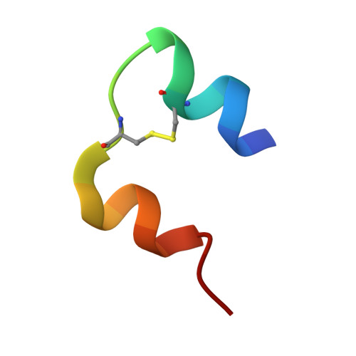
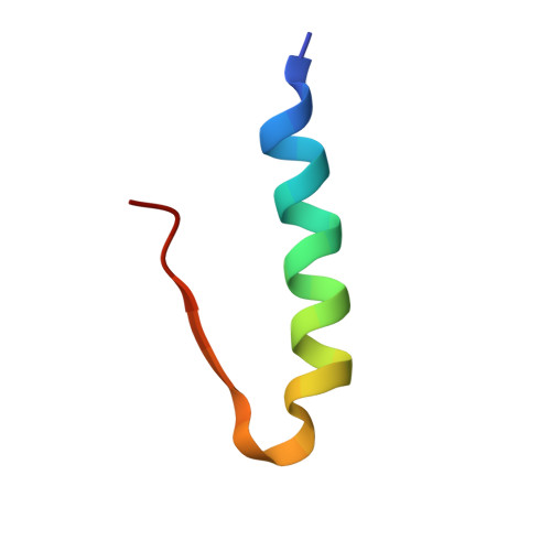
    Insulin is a therapeutic protein that is widely used for the treatment of diabetes. Its biological function was discovered more than 80 years ago and it has since then been characterized extensively. Crystallization of the insulin molecule has always been a key activity since the protein is often administered by subcutaneous injections of crystalline insulin formulations. Over the years, insulin has been crystallized and characterized in a number of crystal systems. Interestingly, we have now discovered two new crystal forms of human insulin. The crystals were obtained when the two chaotropic agents, urea and thiocyanate were present in the crystallization experiments, and their structures were determined by X-ray crystallography. The crystals belong to the orthorhombic and monoclinic crystal systems, with space groups C2221 and C2 respectively. The orthorhombic crystals were obtained at pH 6.5 and contained three insulin hexamers in R6 conformation in the asymmetric unit whilst the monoclinic C2 crystals were obtained at pH 7.0 and contained one R6 hexamer in the asymmetric unit. Common for the two new crystals is a hexamer-hexamer interaction that has not been found in any of the previous crystal forms of insulin. The contacts involve a tight glutamate-glutamate interaction with a distance of 2.3 A between groups. The short distance suggests a low barrier hydrogen bond. In addition, two tyrosine-tyrosine interactions occupying a known phenol binding pocket contribute to the stabilization of the contacts. Within the crystals, distinct binding sites for urea were found, adding further to the discussion on the role of urea in protein denaturation. The change in space group from C2221 to C2 was primarily caused by an increase in pH. The fewer number of hexamer-hexamer interactions comprising the short hydrogen bond in the C2 space group suggest that pH is the driving force. In addition, the distance between the two glutamates increases from 2.32 A in the C2221 crystals to 2.4 A in the C2 crystals. However, in both cases the low barrier hydrogen bond and the tyrosine-tyrosine interaction should contribute to the stability of the crystals which is crucial when used in pharmaceutical formulations.


  • Organizational Affiliation

    Diabetes Protein Engineering, Novo Nordisk A/S, Novo Nordisk Park, DK-2760 Måløv, Denmark. mtno@novonordisk.com


Macromolecules

Find similar proteins by:  Sequence   |   3D Structure  

Entity ID: 1
MoleculeChains Sequence LengthOrganismDetailsImage
Insulin A chain21Homo sapiensMutation(s): 0 
UniProt & NIH Common Fund Data Resources
Find proteins for P01308 (Homo sapiens)
Explore P01308 
Go to UniProtKB:  P01308
PHAROS:  P01308
GTEx:  ENSG00000254647 
Entity Groups  
Sequence Clusters30% Identity50% Identity70% Identity90% Identity95% Identity100% Identity
UniProt GroupP01308
Sequence Annotations
Expand
  • Reference Sequence
Find similar proteins by:  (by identity cutoff)  |  3D Structure
Entity ID: 2
MoleculeChains Sequence LengthOrganismDetailsImage
Insulin B chain30Homo sapiensMutation(s): 0 
UniProt & NIH Common Fund Data Resources
Find proteins for P01308 (Homo sapiens)
Explore P01308 
Go to UniProtKB:  P01308
PHAROS:  P01308
GTEx:  ENSG00000254647 
Entity Groups  
Sequence Clusters30% Identity50% Identity70% Identity90% Identity95% Identity100% Identity
UniProt GroupP01308
Sequence Annotations
Expand
  • Reference Sequence
Small Molecules
Ligands 4 Unique
IDChains Name / Formula / InChI Key2D Diagram3D Interactions
RCO
Query on RCO

Download Ideal Coordinates CCD File 
AB [auth Q]
EB [auth S]
GB [auth U]
IB [auth X]
KA [auth A]
AB [auth Q],
EB [auth S],
GB [auth U],
IB [auth X],
KA [auth A],
LB [auth 1],
NB [auth 3],
OA [auth C],
OB [auth a],
RB [auth c],
TA [auth E],
TB [auth e],
UB [auth g],
VA [auth G],
XA [auth I],
YB [auth i],
ZA [auth K],
ZB [auth k]
RESORCINOL
C6 H6 O2
GHMLBKRAJCXXBS-UHFFFAOYSA-N
ZN
Query on ZN

Download Ideal Coordinates CCD File 
CB [auth R]
JB [auth Y]
MA [auth B]
PB [auth b]
QA [auth D]
CB [auth R],
JB [auth Y],
MA [auth B],
PB [auth b],
QA [auth D],
WB [auth h]
ZINC ION
Zn
PTFCDOFLOPIGGS-UHFFFAOYSA-N
URE
Query on URE

Download Ideal Coordinates CCD File 
BB [auth Q]
FB [auth S]
HB [auth U]
LA [auth A]
MB [auth 1]
BB [auth Q],
FB [auth S],
HB [auth U],
LA [auth A],
MB [auth 1],
PA [auth C],
SA [auth D],
SB [auth d],
UA [auth E],
VB [auth g],
WA [auth G],
YA [auth I]
UREA
C H4 N2 O
XSQUKJJJFZCRTK-UHFFFAOYSA-N
CL
Query on CL

Download Ideal Coordinates CCD File 
DB [auth R]
KB [auth Y]
NA [auth B]
QB [auth b]
RA [auth D]
DB [auth R],
KB [auth Y],
NA [auth B],
QB [auth b],
RA [auth D],
XB [auth h]
CHLORIDE ION
Cl
VEXZGXHMUGYJMC-UHFFFAOYSA-M
Experimental Data & Validation

Experimental Data

  • Method: X-RAY DIFFRACTION
  • Resolution: 2.05 Å
  • R-Value Free: 0.227 
  • R-Value Work: 0.184 
  • R-Value Observed: 0.186 
  • Space Group: C 2 2 21
Unit Cell:
Length ( Å )Angle ( ˚ )
a = 58.936α = 90
b = 219.318β = 90
c = 223.674γ = 90
Software Package:
Software NamePurpose
REFMACrefinement
XDSdata reduction
XSCALEdata scaling
MOLREPphasing

Structure Validation

View Full Validation Report


Entry History 

Deposition Data

Revision History  (Full details and data files)

  • Version 1.0: 2007-12-04
    Type: Initial release
  • Version 1.1: 2011-07-13
    Changes: Advisory, Derived calculations, Version format compliance
  • Version 1.2: 2018-03-07
    Changes: Data collection
  • Version 1.3: 2023-12-27
    Changes: Data collection, Database references, Derived calculations
  • Version 1.4: 2024-04-03
    Changes: Refinement description