2OKV

c-Myc DNA Unwinding Element Binding Protein


Experimental Data Snapshot

  • Method: X-RAY DIFFRACTION
  • Resolution: 2.00 Å
  • R-Value Free: 0.237 
  • R-Value Work: 0.205 
  • R-Value Observed: 0.206 

wwPDB Validation   3D Report Full Report


This is version 1.3 of the entry. See complete history


Literature

Structure and Function of the c-myc DNA-unwinding Element-binding Protein DUE-B.

Kemp, M.Bae, B.Yu, J.P.Ghosh, M.Leffak, M.Nair, S.K.

(2007) J Biol Chem 282: 10441-10448

  • DOI: https://doi.org/10.1074/jbc.M609632200
  • Primary Citation of Related Structures:  
    2OKV

  • PubMed Abstract: 

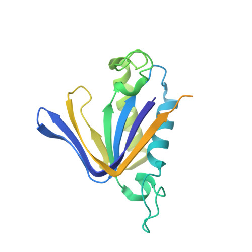
    Local zones of easily unwound DNA are characteristic of prokaryotic and eukaryotic replication origins. The DNA-unwinding element of the human c-myc replication origin is essential for replicator activity and is a target of the DNA-unwinding element-binding protein DUE-B in vivo. We present here the 2.0A crystal structure of DUE-B and complementary biochemical characterization of its biological activity. The structure corresponds to a dimer of the N-terminal domain of the full-length protein and contains many of the structural elements of the nucleotide binding fold. A single magnesium ion resides in the putative active site cavity, which could serve to facilitate ATP hydrolytic activity of this protein. The structure also demonstrates a notable similarity to those of tRNA-editing enzymes. Consistent with this structural homology, the N-terminal core of DUE-B is shown to display both D-aminoacyl-tRNA deacylase activity and ATPase activity. We further demonstrate that the C-terminal portion of the enzyme is disordered and not essential for dimerization. However, this region is essential for DNA binding in vitro and becomes ordered in the presence of DNA.


  • Organizational Affiliation

    Department of Biochemistry and Molecular Biology, Boonshoft School of Medicine, Wright State University, Dayton, Ohio 45435, USA.


Macromolecules
Find similar proteins by:  (by identity cutoff)  |  3D Structure
Entity ID: 1
MoleculeChains Sequence LengthOrganismDetailsImage
Probable D-tyrosyl-tRNA(Tyr) deacylase 1
A, B, C, D
209Homo sapiensMutation(s): 0 
Gene Names: HARS2C20orf88
EC: 3.1
UniProt & NIH Common Fund Data Resources
Find proteins for Q8TEA8 (Homo sapiens)
Explore Q8TEA8 
Go to UniProtKB:  Q8TEA8
PHAROS:  Q8TEA8
GTEx:  ENSG00000125821 
Entity Groups  
Sequence Clusters30% Identity50% Identity70% Identity90% Identity95% Identity100% Identity
UniProt GroupQ8TEA8
Sequence Annotations
Expand
  • Reference Sequence
Experimental Data & Validation

Experimental Data

  • Method: X-RAY DIFFRACTION
  • Resolution: 2.00 Å
  • R-Value Free: 0.237 
  • R-Value Work: 0.205 
  • R-Value Observed: 0.206 
  • Space Group: P 31
Unit Cell:
Length ( Å )Angle ( ˚ )
a = 77.813α = 90
b = 77.813β = 90
c = 106.423γ = 120
Software Package:
Software NamePurpose
REFMACrefinement
ADSCdata collection
HKL-2000data reduction
HKL-2000data scaling
SOLVEphasing

Structure Validation

View Full Validation Report


Entry History 

Deposition Data

Revision History  (Full details and data files)

  • Version 1.0: 2007-01-30
    Type: Initial release
  • Version 1.1: 2008-05-01
    Changes: Version format compliance
  • Version 1.2: 2011-07-13
    Changes: Advisory, Version format compliance
  • Version 1.3: 2023-12-27
    Changes: Data collection, Database references, Derived calculations