2OFF

The crystal structure of Glycogen Phosphorylase b in complex with a potent allosteric inhibitor


Experimental Data Snapshot

  • Method: X-RAY DIFFRACTION
  • Resolution: 2.20 Å
  • R-Value Free: 0.213 
  • R-Value Work: 0.184 
  • R-Value Observed: 0.185 

wwPDB Validation   3D Report Full Report


This is version 1.2 of the entry. See complete history


Literature

FR258900, a potential anti-hyperglycemic drug, binds at the allosteric site of glycogen phosphorylase

Tiraidis, C.Alexacou, K.-M.Zographos, S.E.Leonidas, D.D.Gimisis, T.Oikonomakos, N.G.

(2007) Protein Sci 16: 1773-1782

  • DOI: https://doi.org/10.1110/ps.072925607
  • Primary Citation of Related Structures:  
    2OFF

  • PubMed Abstract: 

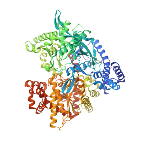
    FR258900 has been discovered as a novel inhibitor of human liver glycogen phosphorylase a and proved to suppress hepatic glycogen breakdown and reduce plasma glucose concentrations in diabetic mice models. To elucidate the mechanism of inhibition, we have determined the crystal structure of the cocrystallized rabbit muscle glycogen phosphorylase b-FR258900 complex and refined it to 2.2 A resolution. The structure demonstrates that the inhibitor binds at the allosteric activator site, where the physiological activator AMP binds. The contacts from FR258900 to glycogen phosphorylase are dominated by nonpolar van der Waals interactions with Gln71, Gln72, Phe196, and Val45' (from the symmetry-related subunit), and also by ionic interactions from the carboxylate groups to the three arginine residues (Arg242, Arg309, and Arg310) that form the allosteric phosphate-recognition subsite. The binding of FR258900 to the protein promotes conformational changes that stabilize an inactive T-state quaternary conformation of the enzyme. The ligand-binding mode is different from those of the potent phenoxy-phthalate and acyl urea inhibitors, previously described, illustrating the broad specificity of the allosteric site.


  • Organizational Affiliation

    Institute of Organic and Pharmaceutical Chemistry, National Hellenic Research Foundation, Athens, Greece.


Macromolecules
Find similar proteins by:  (by identity cutoff)  |  3D Structure
Entity ID: 1
MoleculeChains Sequence LengthOrganismDetailsImage
Glycogen phosphorylase, muscle form842Oryctolagus cuniculusMutation(s): 0 
EC: 2.4.1.1
UniProt
Find proteins for P00489 (Oryctolagus cuniculus)
Explore P00489 
Go to UniProtKB:  P00489
Entity Groups  
Sequence Clusters30% Identity50% Identity70% Identity90% Identity95% Identity100% Identity
UniProt GroupP00489
Sequence Annotations
Expand
  • Reference Sequence
Small Molecules
Ligands 1 Unique
IDChains Name / Formula / InChI Key2D Diagram3D Interactions
OFF
Query on OFF

Download Ideal Coordinates CCD File 
B [auth A]2-DEOXY-3,4-BIS-O-[3-(4-HYDROXYPHENYL)PROPANOYL]-L-THREO-PENTARIC ACID
C23 H20 O10
VJLMRHSHSNLOGC-NOPZTHQXSA-N
Modified Residues  1 Unique
IDChains TypeFormula2D DiagramParent
LLP
Query on LLP
A
L-PEPTIDE LINKINGC14 H22 N3 O7 PLYS
Binding Affinity Annotations 
IDSourceBinding Affinity
OFF BindingDB:  2OFF Ki: 460 (nM) from 1 assay(s)
Binding MOAD:  2OFF Ki: 460 (nM) from 1 assay(s)
PDBBind:  2OFF IC50: 300 (nM) from 1 assay(s)
Experimental Data & Validation

Experimental Data

  • Method: X-RAY DIFFRACTION
  • Resolution: 2.20 Å
  • R-Value Free: 0.213 
  • R-Value Work: 0.184 
  • R-Value Observed: 0.185 
  • Space Group: P 43 21 2
Unit Cell:
Length ( Å )Angle ( ˚ )
a = 128.78α = 90
b = 128.78β = 90
c = 116.051γ = 90
Software Package:
Software NamePurpose
REFMACrefinement
ADSCdata collection
DENZOdata reduction
SCALEPACKdata scaling

Structure Validation

View Full Validation Report


Entry History 

Deposition Data

Revision History  (Full details and data files)

  • Version 1.0: 2007-08-07
    Type: Initial release
  • Version 1.1: 2011-07-13
    Changes: Derived calculations, Version format compliance
  • Version 1.2: 2023-12-27
    Changes: Data collection, Database references, Derived calculations