High-Resolution Crystal Structure of Plant Carboxylesterase AeCXE1, from Actinidia eriantha, and Its Complex with a High-Affinity Inhibitor Paraoxon.
Ileperuma, N.R., Marshall, S.D., Squire, C.J., Baker, H.M., Oakeshott, J.G., Russell, R.J., Plummer, K.M., Newcomb, R.D., Baker, E.N.(2007) Biochemistry 46: 1851-1859
- PubMed: 17256879
- DOI: https://doi.org/10.1021/bi062046w
- Primary Citation of Related Structures:
2O7R, 2O7V - PubMed Abstract:
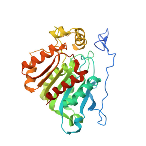
Carboxylesterases (CXEs) are widely distributed in plants, where they have been implicated in roles that include plant defense, plant development, and secondary metabolism. We have cloned, overexpressed, purified, and crystallized a carboxylesterase from the kiwifruit species Actinidia eriantha (AeCXE1). The structure of AeCXE1 was determined by X-ray crystallography at 1.4 A resolution. The crystal structure revealed that AeCXE1 is a member of the alpha/beta-hydrolase fold superfamily, most closely related structurally to the hormone-sensitive lipase subgroup. The active site of the enzyme, located in an 11 A deep hydrophobic gorge, contains the conserved catalytic triad residues Ser169, Asp276, and His306. Kinetic analysis using artificial ester substrates showed that the enzyme can hydrolyze a range of carboxylester substrates with acyl groups ranging from C2 to C16, with a preference for butyryl moieties. This preference was supported by the discovery of a three-carbon acyl adduct bound to the active site Ser169 in the native structure. AeCXE1 was also found to be inhibited by organophosphates, with paraoxon (IC50 = 1.1 muM) a more potent inhibitor than dimethylchlorophosphate (DMCP; IC50 = 9.2 muM). The structure of AeCXE1 with paraoxon bound was determined at 2.3 A resolution and revealed that the inhibitor binds covalently to the catalytic serine residue, with virtually no change in the structure of the enzyme. The structural information for AeCXE1 provides a basis for addressing the wider functional roles of carboxylesterases in plants.
- School of Biological Sciences, University of Auckland, Auckland, New Zealand.
Organizational Affiliation: