2O4F

Structure of a parallel-stranded guanine tetraplex crystallised with monovalent ions


Experimental Data Snapshot

  • Method: X-RAY DIFFRACTION
  • Resolution: 1.50 Å
  • R-Value Free: 0.257 
  • R-Value Work: 0.204 
  • R-Value Observed: 0.206 

wwPDB Validation   3D Report Full Report


This is version 1.3 of the entry. See complete history


Literature

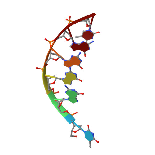
Structure of a d(TGGGGT) quadruplex crystallized in the presence of Li+ ions.

Creze, C.Rinaldi, B.Haser, R.Bouvet, P.Gouet, P.

(2007) Acta Crystallogr D Biol Crystallogr 63: 682-688

  • DOI: https://doi.org/10.1107/S0907444907013315
  • Primary Citation of Related Structures:  
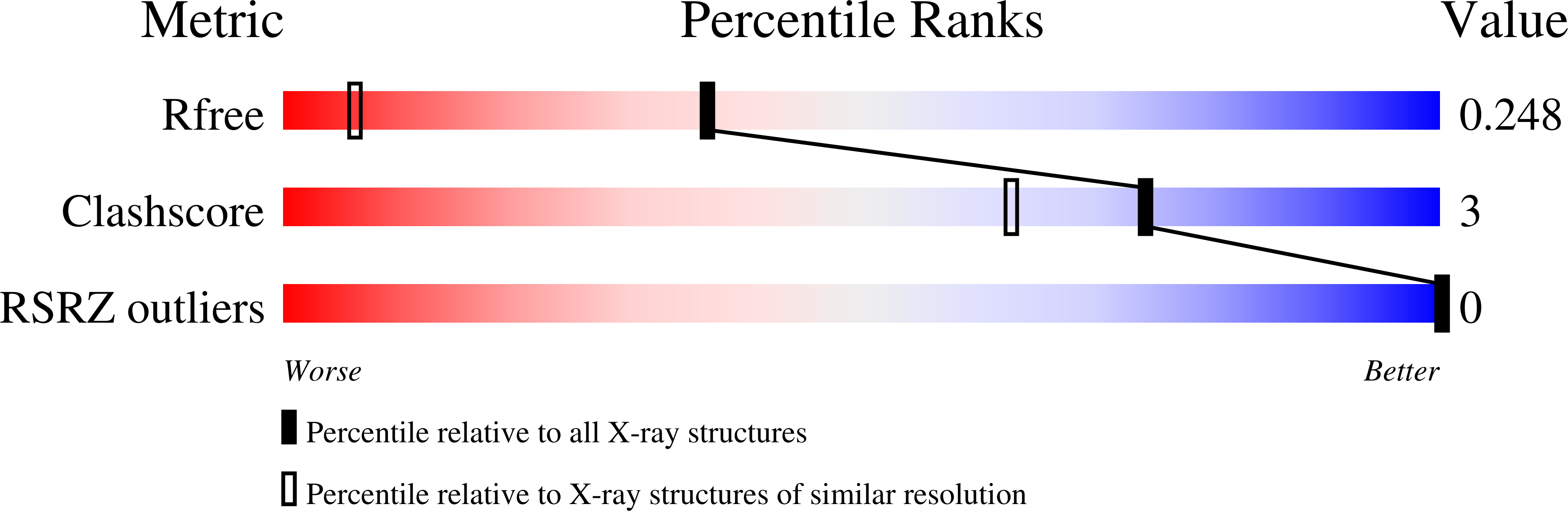
    2O4F

  • PubMed Abstract: 

    A parallel 5'-d(TGGGGT)-3' quadruplex was formed in Na(+) solution and crystallized using lithium sulfate as the main precipitating agent. The X-ray structure was determined to 1.5 A resolution in space group P2(1) by molecular replacement. The asymmetric unit consists of a characteristic motif of two quadruplexes stacked at their 5' ends. All nucleotides are clearly defined in the density and could be positioned. A single bound Li(+) ion is observed at the surface of the column formed by the two joined molecules. Thus, this small alkali metal ion appears to be unsuitable as a replacement for the Na(+) ion in the central channel of G-quartets, unlike K(+) or Tl(+) ions. A well conserved constellation of water molecules is observed in the grooves of the dimeric structure.


  • Organizational Affiliation

    Laboratoire de BioCristallographie, Institut de Biologie et Chimie des Protéines, CNRS-UCBL, UMR 5086, IFR128 BioSciences Lyon-Gerland, 7 Passage du Vercors, 69007 Lyon, France.


Macromolecules

Find similar nucleic acids by:  Sequence   |   3D Structure  

Entity ID: 1
MoleculeChains LengthOrganismImage
5'-D(*TP*GP*GP*GP*GP*T)-3'
A, B, C, D, E
A, B, C, D, E, F, G, H
6N/A
Sequence Annotations
Expand
  • Reference Sequence
Experimental Data & Validation

Experimental Data

  • Method: X-RAY DIFFRACTION
  • Resolution: 1.50 Å
  • R-Value Free: 0.257 
  • R-Value Work: 0.204 
  • R-Value Observed: 0.206 
  • Space Group: P 1 21 1
Unit Cell:
Length ( Å )Angle ( ˚ )
a = 28.92α = 90
b = 57.37β = 108
c = 35.92γ = 90
Software Package:
Software NamePurpose
CNSrefinement
PDB_EXTRACTdata extraction
ProDCdata collection
XDSdata reduction
XSCALEdata scaling
AMoREphasing

Structure Validation

View Full Validation Report


Entry History 

Deposition Data

Revision History  (Full details and data files)

  • Version 1.0: 2007-05-22
    Type: Initial release
  • Version 1.1: 2007-10-30
    Changes: Version format compliance
  • Version 1.2: 2011-07-13
    Changes: Version format compliance
  • Version 1.3: 2023-08-30
    Changes: Data collection, Database references, Derived calculations, Refinement description