


Comparison of MAS solid-state NMR and X-ray crystallographic data in the analysis of protein dynamics in the solid state
Agarwal, V., Faelber, K., Hologne, M., Chevelkov, V., Oschkinat, H., Diehl, A., Reif, B.To be published.
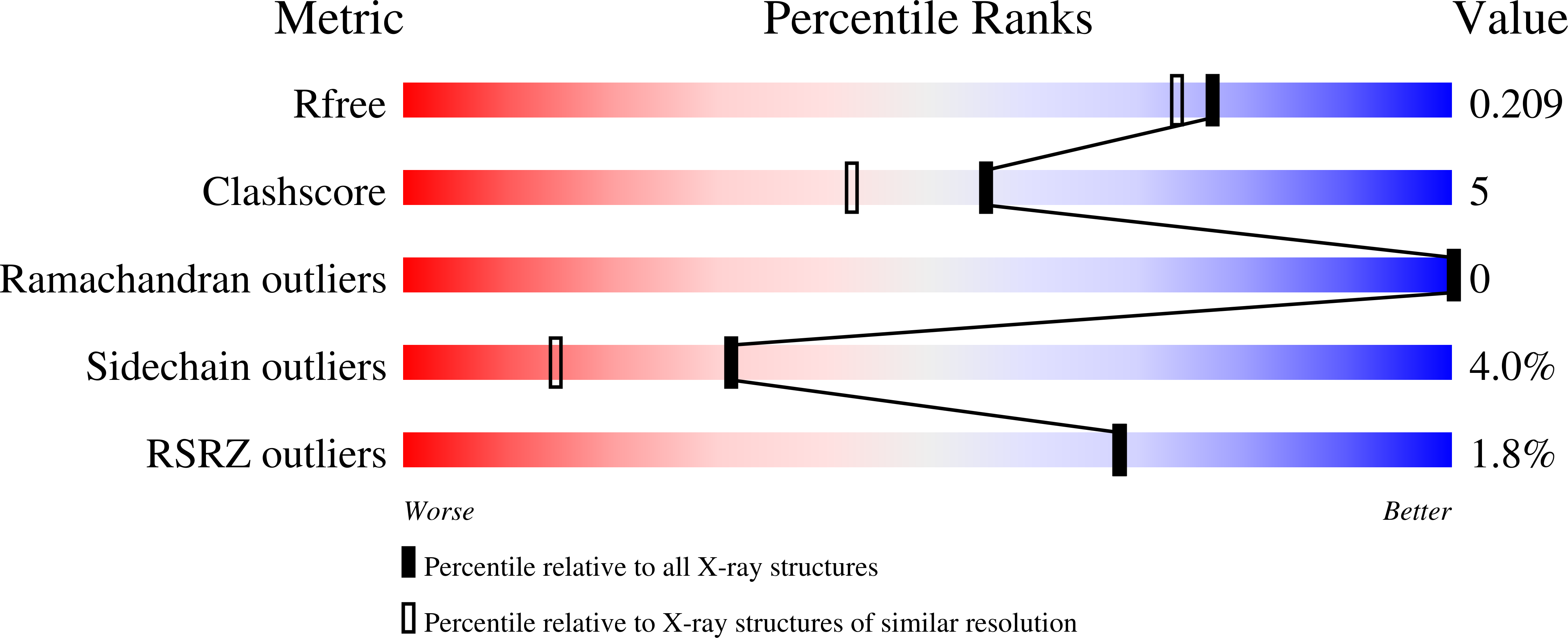
Experimental Data Snapshot
wwPDB Validation 3D Report Full Report
Entity ID: 1 | |||||
|---|---|---|---|---|---|
| Molecule | Chains | Sequence Length | Organism | Details | Image |
| Spectrin alpha chain, brain | 62 | Gallus gallus | Mutation(s): 0 | 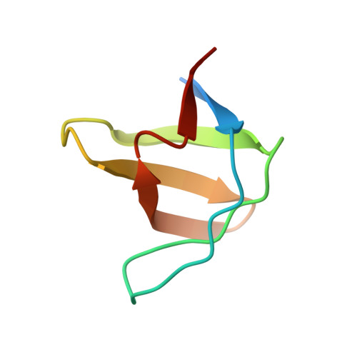 | |
UniProt | |||||
Find proteins for P07751 (Gallus gallus) Explore P07751 Go to UniProtKB: P07751 | |||||
Entity Groups | |||||
| Sequence Clusters | 30% Identity50% Identity70% Identity90% Identity95% Identity100% Identity | ||||
| UniProt Group | P07751 | ||||
Sequence AnnotationsExpand | |||||
| |||||
| Length ( Å ) | Angle ( ˚ ) |
|---|---|
| a = 34.465 | α = 90 |
| b = 42.479 | β = 90 |
| c = 50.799 | γ = 90 |
| Software Name | Purpose |
|---|---|
| REFMAC | refinement |
| MAR345 | data collection |
| XDS | data reduction |
| XDS | data scaling |
| AMoRE | phasing |
RCSB PDB (citation) is hosted by
RCSB PDB is a member of the

