2MTA

CRYSTAL STRUCTURE OF A TERNARY ELECTRON TRANSFER COMPLEX BETWEEN METHYLAMINE DEHYDROGENASE, AMICYANIN AND A C-TYPE CYTOCHROME


Experimental Data Snapshot

  • Method: X-RAY DIFFRACTION
  • Resolution: 2.40 Å
  • R-Value Work: 0.179 
  • R-Value Observed: 0.179 

wwPDB Validation   3D Report Full Report


This is version 2.0 of the entry. See complete history


Literature

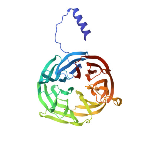
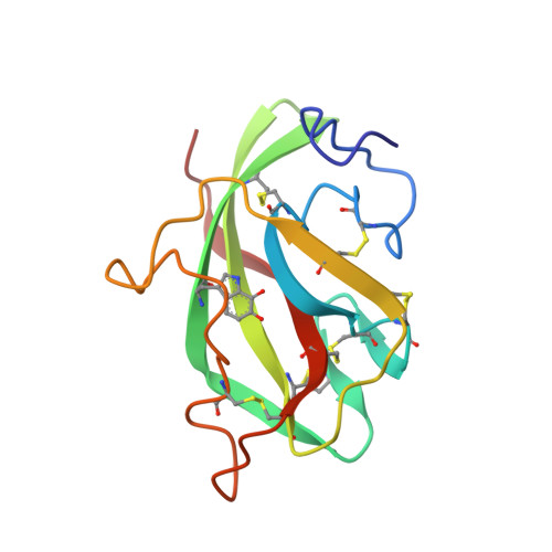
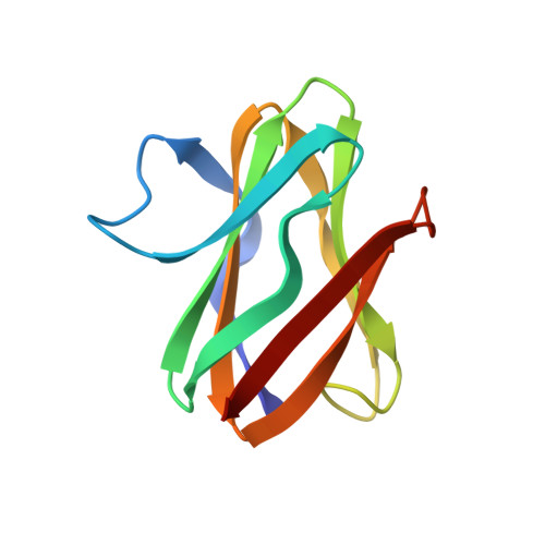
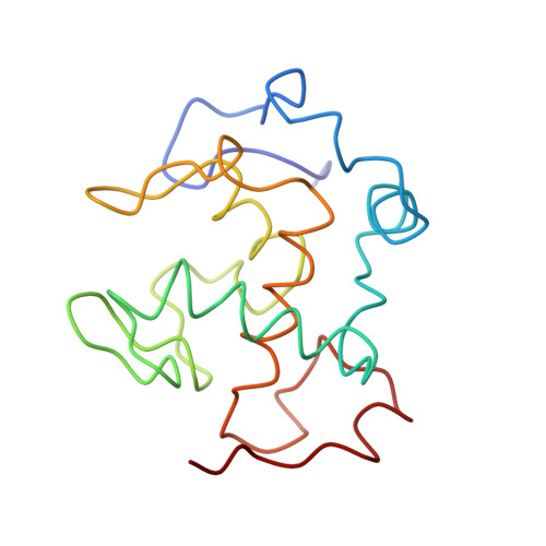
Structure of an electron transfer complex: methylamine dehydrogenase, amicyanin, and cytochrome c551i.

Chen, L.Durley, R.C.Mathews, F.S.Davidson, V.L.

(1994) Science 264: 86-90

  • DOI: https://doi.org/10.1126/science.8140419
  • Primary Citation of Related Structures:  
    2MTA

  • PubMed Abstract: 

    The crystal structure of a ternary protein complex has been determined at 2.4 angstrom resolution. The complex is composed of three electron transfer proteins from Paracoccus denitrificans, the quinoprotein methylamine dehydrogenase, the blue copper protein amicyanin, and the cytochrome c551i. The central region of the c551i is folded similarly to several small bacterial c-type cytochromes; there is a 45-residue extension at the amino terminus and a 25-residue extension at the carboxyl terminus. The methylamine dehydrogenase-amicyanin interface is largely hydrophobic, whereas the amicyanin-cytochrome interface is more polar, with several charged groups present on each surface. Analysis of the simplest electron transfer pathways between the redox partners points out the importance of other factors such as energetics in determining the electron transfer rates.


  • Organizational Affiliation

    Department of Biochemistry and Molecular Biophysics, Washington University School of Medicine, St. Louis, MO 63110.


Macromolecules
Find similar proteins by:  (by identity cutoff)  |  3D Structure
Entity ID: 1
MoleculeChains Sequence LengthOrganismDetailsImage
METHYLAMINE DEHYDROGENASE (HEAVY SUBUNIT)A [auth H]373Paracoccus denitrificansMutation(s): 0 
EC: 1.4.99.3
UniProt
Find proteins for P29894 (Paracoccus denitrificans)
Explore P29894 
Go to UniProtKB:  P29894
Entity Groups  
Sequence Clusters30% Identity50% Identity70% Identity90% Identity95% Identity100% Identity
UniProt GroupP29894
Sequence Annotations
Expand
  • Reference Sequence
Find similar proteins by:  (by identity cutoff)  |  3D Structure
Entity ID: 2
MoleculeChains Sequence LengthOrganismDetailsImage
METHYLAMINE DEHYDROGENASE (LIGHT SUBUNIT)B [auth L]125Paracoccus denitrificansMutation(s): 0 
EC: 1.4.99.3
UniProt
Find proteins for P22619 (Paracoccus denitrificans)
Explore P22619 
Go to UniProtKB:  P22619
Entity Groups  
Sequence Clusters30% Identity50% Identity70% Identity90% Identity95% Identity100% Identity
UniProt GroupP22619
Sequence Annotations
Expand
  • Reference Sequence
Find similar proteins by:  (by identity cutoff)  |  3D Structure
Entity ID: 3
MoleculeChains Sequence LengthOrganismDetailsImage
AMICYANINC [auth A]105Paracoccus denitrificansMutation(s): 0 
UniProt
Find proteins for P22364 (Paracoccus denitrificans)
Explore P22364 
Go to UniProtKB:  P22364
Entity Groups  
Sequence Clusters30% Identity50% Identity70% Identity90% Identity95% Identity100% Identity
UniProt GroupP22364
Sequence Annotations
Expand
  • Reference Sequence
Find similar proteins by:  (by identity cutoff)  |  3D Structure
Entity ID: 4
MoleculeChains Sequence LengthOrganismDetailsImage
CYTOCHROME C551ID [auth C]147Paracoccus denitrificansMutation(s): 0 
UniProt
Find proteins for P29899 (Paracoccus denitrificans)
Explore P29899 
Go to UniProtKB:  P29899
Entity Groups  
Sequence Clusters30% Identity50% Identity70% Identity90% Identity95% Identity100% Identity
UniProt GroupP29899
Sequence Annotations
Expand
  • Reference Sequence
Small Molecules
Ligands 3 Unique
IDChains Name / Formula / InChI Key2D Diagram3D Interactions
HEC
Query on HEC

Download Ideal Coordinates CCD File 
G [auth C]HEME C
C34 H34 Fe N4 O4
HXQIYSLZKNYNMH-LJNAALQVSA-N
PO4
Query on PO4

Download Ideal Coordinates CCD File 
E [auth H]PHOSPHATE ION
O4 P
NBIIXXVUZAFLBC-UHFFFAOYSA-K
CU
Query on CU

Download Ideal Coordinates CCD File 
F [auth A]COPPER (II) ION
Cu
JPVYNHNXODAKFH-UHFFFAOYSA-N
Modified Residues  1 Unique
IDChains TypeFormula2D DiagramParent
TRQ
Query on TRQ
B [auth L]L-PEPTIDE LINKINGC11 H10 N2 O4TRP
Experimental Data & Validation

Experimental Data

  • Method: X-RAY DIFFRACTION
  • Resolution: 2.40 Å
  • R-Value Work: 0.179 
  • R-Value Observed: 0.179 
  • Space Group: C 2 2 21
Unit Cell:
Length ( Å )Angle ( ˚ )
a = 148.81α = 90
b = 68.85β = 90
c = 187.18γ = 90
Software Package:
Software NamePurpose
X-PLORmodel building
X-PLORrefinement
X-PLORphasing

Structure Validation

View Full Validation Report


Entry History 

Deposition Data

Revision History  (Full details and data files)

  • Version 1.0: 1994-01-31
    Type: Initial release
  • Version 1.1: 2008-03-03
    Changes: Version format compliance
  • Version 1.2: 2011-07-13
    Changes: Version format compliance
  • Version 2.0: 2021-03-10
    Changes: Advisory, Atomic model, Data collection, Database references, Derived calculations, Non-polymer description, Other, Structure summary