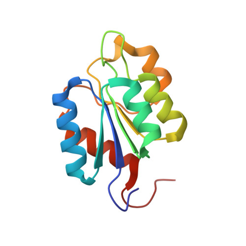
Ligand-induced folding of a two-component signaling receiver domain.
Ocasio, V.J., Correa, F., Gardner, K.H.(2015) Biochemistry 54: 1353-1363
- PubMed: 25629646
- DOI: https://doi.org/10.1021/bi501143b
- Primary Citation of Related Structures:
2MSW - PubMed Abstract:
To survive and adapt to environmental changes, bacteria commonly use two-component signaling systems. Minimally, these pathways use histidine kinases (HKs) to detect environmental signals, harnessing these to control phosphorylation levels of receiver (REC) domains of downstream response regulators that convert this signal into physiological responses. Studies of several prototypical REC domains suggest that phosphorylation shifts these proteins between inactive and active structures that are globally similar and well-folded. However, it is unclear how globally these findings hold within REC domains in general, particularly when they are considered within full-length proteins. Here, we present EL_LovR, a full-length REC-only protein that is phosphorylated in response to blue light in the marine α-proteobacterium, Erythrobacter litoralis HTCC2594. Notably, EL_LovR is similar to comparable REC-only proteins used in bacterial general stress responses, where genetic evidence suggests that their potent phosphatase activity is important to shut off such systems. Size exclusion chromatography, light scattering, and solution NMR experiments show that EL_LovR is monomeric and unfolded in solution under conditions routinely used for other REC structure determinations. Addition of Mg(2+) and phosphorylation induce progressively greater degrees of tertiary structure stabilization, with the solution structure of the fully activated EL_LovR adopting the canonical receiver domain fold. Parallel functional assays show that EL_LovR has a fast dephosphorylation rate, consistent with its proposed function as a phosphate sink that depletes the HK phosphoryl group, promoting the phosphatase activity of this enzyme. Our findings demonstrate that EL_LovR undergoes substantial ligand-dependent conformational changes that have not been reported for other RRs, expanding the scope of conformational changes and regulation used by REC domains, critical components of bacterial signaling systems.
Organizational Affiliation:
Departments of Biophysics and Biochemistry, UT Southwestern Medical Center , Dallas, Texas 75390-8816, United States.