2MKP

N domain of cardiac troponin C bound to the switch fragment of fast skeletal troponin I at pH 6


Experimental Data Snapshot

  • Method: SOLUTION NMR
  • Conformers Calculated: 50 
  • Conformers Submitted: 10 
  • Selection Criteria: structures with the least restraint violations 

wwPDB Validation   3D Report Full Report


This is version 1.1 of the entry. See complete history


Literature

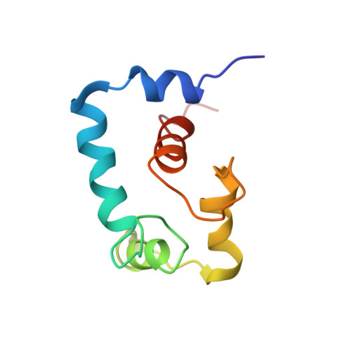
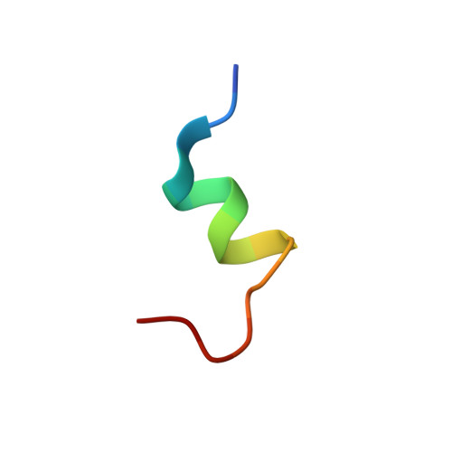
Conformation of the critical pH sensitive region of troponin depends upon a single residue in troponin I.

Robertson, I.M.Pineda-Sanabria, S.E.Holmes, P.C.Sykes, B.D.

(2014) Arch Biochem Biophys 552-553: 40-49

  • DOI: https://doi.org/10.1016/j.abb.2013.12.003
  • Primary Citation of Related Structures:  
    2MKP

  • PubMed Abstract: 

    The calcium sensitivity of cardiac and skeletal muscle is reduced during cytosolic acidosis, and this inhibition is more pronounced in cardiac muscle. Replacing cardiac troponin I with skeletal troponin I reduces the pH sensitivity of cardiac muscle. This diminished pH sensitivity depends on a single amino acid difference in troponin I: an alanine in cardiac and a histidine in skeletal. Studies suggested that when this histidine is protonated, it forms an electrostatic interaction with glutamate 19 on the surface of cardiac troponin C. Structures of the skeletal and cardiac troponin complexes show very different conformations for the region of troponin I surrounding this residue. In this study, we determined the structure of skeletal troponin I bound to cardiac troponin C. Skeletal troponin I is found to bind to cardiac troponin C with histidine 130 in close proximity to glutamate 19. This conformation is homologous to the crystal structure of the skeletal troponin complex; but different than in the cardiac complex. We show that an A162H variant of cardiac troponin I adopts a conformation similar to the skeletal structure. The implications of these structural differences in the context of cardiac muscle regulation are discussed.


  • Organizational Affiliation

    Department of Biochemistry, University of Alberta, 4-19 Medical Sciences Building, Edmonton, Alberta T6G 2H7, Canada.


Macromolecules
Find similar proteins by:  (by identity cutoff)  |  3D Structure
Entity ID: 1
MoleculeChains Sequence LengthOrganismDetailsImage
Troponin C, slow skeletal and cardiac musclesA [auth C]89Homo sapiensMutation(s): 0 
Gene Names: TNNC1TNNC
UniProt & NIH Common Fund Data Resources
Find proteins for P63316 (Homo sapiens)
Explore P63316 
Go to UniProtKB:  P63316
PHAROS:  P63316
GTEx:  ENSG00000114854 
Entity Groups  
Sequence Clusters30% Identity50% Identity70% Identity90% Identity95% Identity100% Identity
UniProt GroupP63316
Sequence Annotations
Expand
  • Reference Sequence

Find similar proteins by:  Sequence   |   3D Structure  

Entity ID: 2
MoleculeChains Sequence LengthOrganismDetailsImage
Troponin I, fast skeletal muscleB [auth I]17Homo sapiensMutation(s): 0 
Gene Names: TNNI2
UniProt & NIH Common Fund Data Resources
Find proteins for P48788 (Homo sapiens)
Explore P48788 
Go to UniProtKB:  P48788
PHAROS:  P48788
GTEx:  ENSG00000130598 
Entity Groups  
Sequence Clusters30% Identity50% Identity70% Identity90% Identity95% Identity100% Identity
UniProt GroupP48788
Sequence Annotations
Expand
  • Reference Sequence
Small Molecules
Ligands 1 Unique
IDChains Name / Formula / InChI Key2D Diagram3D Interactions
CA
Query on CA

Download Ideal Coordinates CCD File 
C
CALCIUM ION
Ca
BHPQYMZQTOCNFJ-UHFFFAOYSA-N
Experimental Data & Validation

Experimental Data

  • Method: SOLUTION NMR
  • Conformers Calculated: 50 
  • Conformers Submitted: 10 
  • Selection Criteria: structures with the least restraint violations 

Structure Validation

View Full Validation Report


Entry History 

Deposition Data

Revision History  (Full details and data files)

  • Version 1.0: 2014-02-26
    Type: Initial release
  • Version 1.1: 2014-06-18
    Changes: Database references