Structure, dynamics, lipid binding, and physiological relevance of the putative GTPase-binding domain of Dictyostelium formin C.
Dames, S.A., Junemann, A., Sass, H.J., Schonichen, A., Stopschinski, B.E., Grzesiek, S., Faix, J., Geyer, M.(2011) J Biol Chem 286: 36907-36920
- PubMed: 21846933
- DOI: https://doi.org/10.1074/jbc.M111.225052
- Primary Citation of Related Structures:
2L1A - PubMed Abstract:
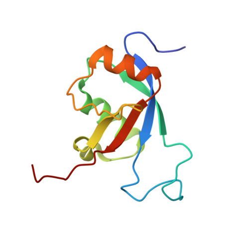
Dictyostelium Formin C (ForC) is involved in the regulation of local actin cytoskeleton reorganization (e.g. during cellular adhesion or migration). ForC contains formin homology 2 and 3 (FH2 and -3) domains and an N-terminal putative GTPase-binding domain (GBD) but lacks a canonical FH1 region. To better understand the role of the GBD, its structure, dynamics, lipid-binding properties, and cellular functions were analyzed by NMR and CD spectroscopy and by in vivo fluorescence microscopy. Moreover, the program CS-Rosetta was tested for the structure prediction based on chemical shift data only. The ForC GBD adopts an ubiquitin-like α/β-roll fold with an unusually long loop between β-strands 1 and 2. Based on the lipid-binding data, the presence of DPC micelles induces the formation of α-helical secondary structure and a rearrangement of the tertiary structure. Lipid-binding studies with a mutant protein and a peptide suggest that the β1-β2 loop is not relevant for these conformational changes. Whereas small amounts of negatively charged phosphoinositides (1,2-dioctanoyl-sn-glycero-3-(phosphoinositol 4,5-bisphosphate) and 1,2-dihexanoyl-sn-glycero-3-(phosphoinositol 3,4,5-trisphosphate)) lower the micelle concentration necessary to induce the observed spectral changes, other negatively charged phospholipids (1,2-dihexanoyl-sn-glycero-3-(phospho-L-serine) and 1,2-dihexanoyl-sn-glycero-3-phospho-(1'-rac-glycerol)) had no such effect. Interestingly, bicelles and micelles composed of diacylphosphocholines had no effect on the GBD structure. Our data suggest a model in which part of the large positively charged surface area of the GBD mediates localization to specific membrane patches, thereby regulating interactions with signaling proteins. Our cellular localization studies show that both the GBD and the FH3 domain are required for ForC targeting to cell-cell contacts and early phagocytic cups and macropinosomes.
Organizational Affiliation:
Department of Chemistry, Technische Universität München, Lichtenbergstrasse 4, 85747 Garching, Germany. sonja.dames@tum.de