2JD9

Structure of a pectin binding carbohydrate binding module determined in an orthorhombic crystal form.


Experimental Data Snapshot

  • Method: X-RAY DIFFRACTION
  • Resolution: 1.80 Å
  • R-Value Free: 0.240 
  • R-Value Work: 0.172 
  • R-Value Observed: 0.175 

wwPDB Validation   3D Report Full Report


This is version 1.2 of the entry. See complete history


Literature

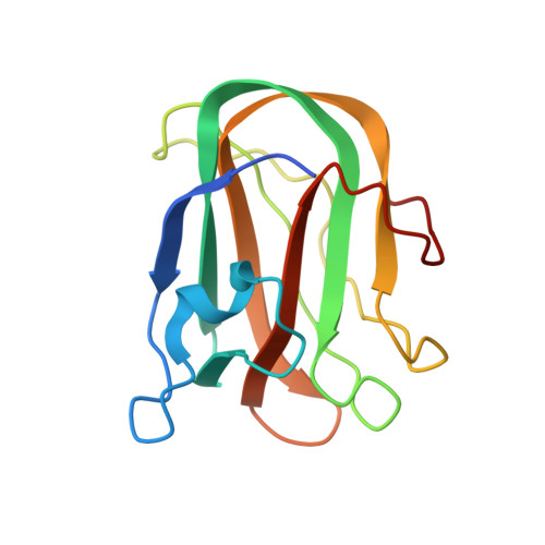
Identification and Characterization of a Novel Periplasmic Polygalacturonic Acid Binding Protein from Yersinia Enterolitica

Abbott, D.W.Hyrnuik, S.Boraston, A.B.

(2007) J Mol Biol 367: 1023

  • DOI: https://doi.org/10.1016/j.jmb.2007.01.030
  • Primary Citation of Related Structures:  
    2JD9, 2JDA

  • PubMed Abstract: 

    A number of bacteria in the family Enterobacteriaceae harbor the genes comprising well-developed pectinolytic pathways (e.g. Erwinia sp.) or abridged versions of this pathway (e.g. Yersinia sp.). One of the most enigmatic components present in some of these pathways is a small gene that encodes a predicted secreted protein of approximately 160 amino acid residues with unknown function. This protein shows distant amino acid sequence similarity over its entire length to galactose-specific family 32 carbohydrate-binding modules (CBMs). Here we demonstrate the ability of the Yersinia enterocolitica example, here called YeCBM32, to bind polygalacturonic acid containing components of pectin. This binding is selective for highly polymerized galacturonic acid and shows a complex mode of polysaccharide recognition. The high resolution X-ray crystal structure (1.35 A) shows YeCBM32s overall structural similarity to galactose specific CBMs and conserved binding site location but reveals a substantially different binding site topology, which likely reflects its unique polymeric and acidic ligand. The results suggest the possibility of a unique role for YeCBM32 in polygalacturonic acid transport.


  • Organizational Affiliation

    Biochemistry and Microbiology, University of Victoria, PO Box 3055 STN CSC, Victoria BC, Canada V8W 3P6.


Macromolecules
Find similar proteins by:  (by identity cutoff)  |  3D Structure
Entity ID: 1
MoleculeChains Sequence LengthOrganismDetailsImage
YECBM32145Yersinia enterocoliticaMutation(s): 0 
UniProt
Find proteins for A1JSS7 (Yersinia enterocolitica serotype O:8 / biotype 1B (strain NCTC 13174 / 8081))
Explore A1JSS7 
Go to UniProtKB:  A1JSS7
Entity Groups  
Sequence Clusters30% Identity50% Identity70% Identity90% Identity95% Identity100% Identity
UniProt GroupA1JSS7
Sequence Annotations
Expand
  • Reference Sequence
Small Molecules
Ligands 1 Unique
IDChains Name / Formula / InChI Key2D Diagram3D Interactions
CA
Query on CA

Download Ideal Coordinates CCD File 
B [auth A]CALCIUM ION
Ca
BHPQYMZQTOCNFJ-UHFFFAOYSA-N
Experimental Data & Validation

Experimental Data

  • Method: X-RAY DIFFRACTION
  • Resolution: 1.80 Å
  • R-Value Free: 0.240 
  • R-Value Work: 0.172 
  • R-Value Observed: 0.175 
  • Space Group: P 21 21 21
Unit Cell:
Length ( Å )Angle ( ˚ )
a = 34.765α = 90
b = 42.63β = 90
c = 98.681γ = 90
Software Package:
Software NamePurpose
REFMACrefinement
MOLREPphasing

Structure Validation

View Full Validation Report


Entry History 

Deposition Data

Revision History  (Full details and data files)

  • Version 1.0: 2007-01-16
    Type: Initial release
  • Version 1.1: 2011-05-08
    Changes: Version format compliance
  • Version 1.2: 2011-07-13
    Changes: Version format compliance