2IGM

Crystal structure of recombinant pyranose 2-oxidase H548N mutant


Experimental Data Snapshot

  • Method: X-RAY DIFFRACTION
  • Resolution: 1.90 Å
  • R-Value Free: 0.189 
  • R-Value Work: 0.152 
  • R-Value Observed: 0.152 

wwPDB Validation   3D Report Full Report


Ligand Structure Quality Assessment 


This is version 1.5 of the entry. See complete history


Literature

Structural basis for substrate binding and regioselective oxidation of monosaccharides at c3 by pyranose 2-oxidase.

Kujawa, M.Ebner, H.Leitner, C.Hallberg, B.M.Prongjit, M.Sucharitakul, J.Ludwig, R.Rudsander, U.Peterbauer, C.Chaiyen, P.Haltrich, D.Divne, C.

(2006) J Biol Chem 281: 35104-35115

  • DOI: https://doi.org/10.1074/jbc.M604718200
  • Primary Citation of Related Structures:  
    2IGK, 2IGM, 2IGN, 2IGO

  • PubMed Abstract: 

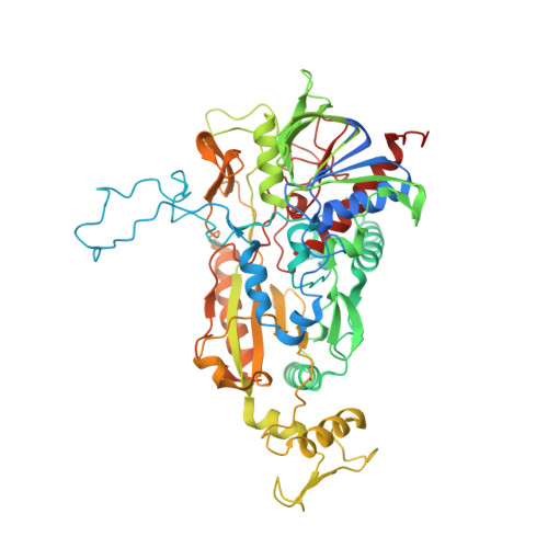
    Pyranose 2-oxidase (P2Ox) participates in fungal lignin degradation by producing the H2O2 needed for lignin-degrading peroxidases. The enzyme oxidizes cellulose- and hemicellulose-derived aldopyranoses at C2 preferentially, but also on C3, to the corresponding ketoaldoses. To investigate the structural determinants of catalysis, covalent flavinylation, substrate binding, and regioselectivity, wild-type and mutant P2Ox enzymes were produced and characterized biochemically and structurally. Removal of the histidyl-FAD linkage resulted in a catalytically competent enzyme containing tightly, but noncovalently bound FAD. This mutant (H167A) is characterized by a 5-fold lower kcat, and a 35-mV lower redox potential, although no significant structural changes were seen in its crystal structure. In previous structures of P2Ox, the substrate loop (residues 452-457) covering the active site has been either disordered or in a conformation incompatible with carbohydrate binding. We present here the crystal structure of H167A in complex with a slow substrate, 2-fluoro-2-deoxy-D-glucose. Based on the details of 2-fluoro-2-deoxy-D-glucose binding in position for oxidation at C3, we also outline a probable binding mode for D-glucose positioned for regioselective oxidation at C2. The tentative determinant for discriminating between the two binding modes is the position of the O6 hydroxyl group, which in the C2-oxidation mode can make favorable interactions with Asp452 in the substrate loop and, possibly, a nearby arginine residue (Arg472). We also substantiate our hypothesis with steady-state kinetics data for the alanine replacements of Asp452 and Arg472 as well as the double alanine 452/472 mutant.


  • Organizational Affiliation

    School of Biotechnology, Royal Institute of Technology, Albanova University Center, SE-106 91 Stockholm, Sweden.


Macromolecules
Find similar proteins by:  (by identity cutoff)  |  3D Structure
Entity ID: 1
MoleculeChains Sequence LengthOrganismDetailsImage
Pyranose oxidase623Trametes ochraceaMutation(s): 1 
Gene Names: p2o
EC: 1.1.3.10
UniProt
Find proteins for Q7ZA32 (Trametes ochracea)
Explore Q7ZA32 
Go to UniProtKB:  Q7ZA32
Entity Groups  
Sequence Clusters30% Identity50% Identity70% Identity90% Identity95% Identity100% Identity
UniProt GroupQ7ZA32
Sequence Annotations
Expand
  • Reference Sequence
Small Molecules
Ligands 2 Unique
IDChains Name / Formula / InChI Key2D Diagram3D Interactions
FAD
Query on FAD

Download Ideal Coordinates CCD File 
I [auth A]
K [auth B]
M [auth D]
O [auth C]
Q [auth E]
I [auth A],
K [auth B],
M [auth D],
O [auth C],
Q [auth E],
S [auth F],
U [auth G],
W [auth H]
FLAVIN-ADENINE DINUCLEOTIDE
C27 H33 N9 O15 P2
VWWQXMAJTJZDQX-UYBVJOGSSA-N
MES
Query on MES

Download Ideal Coordinates CCD File 
J [auth A]
L [auth B]
N [auth D]
P [auth C]
R [auth E]
J [auth A],
L [auth B],
N [auth D],
P [auth C],
R [auth E],
T [auth F],
V [auth G],
X [auth H]
2-(N-MORPHOLINO)-ETHANESULFONIC ACID
C6 H13 N O4 S
SXGZJKUKBWWHRA-UHFFFAOYSA-N
Experimental Data & Validation

Experimental Data

  • Method: X-RAY DIFFRACTION
  • Resolution: 1.90 Å
  • R-Value Free: 0.189 
  • R-Value Work: 0.152 
  • R-Value Observed: 0.152 
  • Space Group: P 1 21 1
Unit Cell:
Length ( Å )Angle ( ˚ )
a = 168.431α = 90
b = 103.142β = 106.08
c = 168.915γ = 90
Software Package:
Software NamePurpose
REFMACrefinement
MAR345data collection
XDSdata reduction
XSCALEdata scaling

Structure Validation

View Full Validation Report


Ligand Structure Quality Assessment 


Entry History 

Deposition Data

  • Released Date: 2006-10-10 
  • Deposition Author(s): Divne, C.

Revision History  (Full details and data files)

  • Version 1.0: 2006-10-10
    Type: Initial release
  • Version 1.1: 2008-05-01
    Changes: Version format compliance
  • Version 1.2: 2011-07-13
    Changes: Version format compliance
  • Version 1.3: 2014-11-12
    Changes: Structure summary
  • Version 1.4: 2017-10-18
    Changes: Refinement description
  • Version 1.5: 2021-10-20
    Changes: Database references, Derived calculations