


Crystal structure of glucosamine-phosphate N-acetyltransferase 1
Wu, H., Min, J., Zeng, H., Loppnau, P., Weigelt, J., Sundstrom, M., Arrowsmith, C.H., Edwards, A.M., Bochkarev, A., Plotnikov, A.N.To be published.
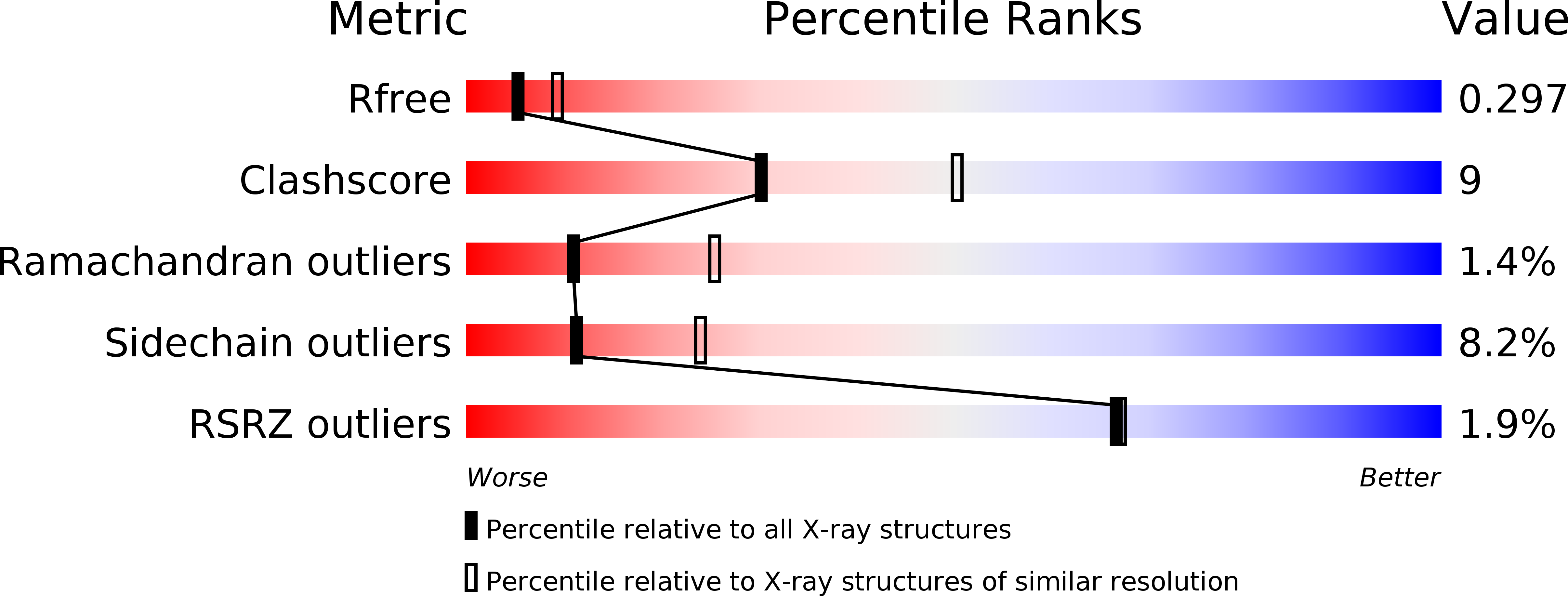
Experimental Data Snapshot
wwPDB Validation 3D Report Full Report
Entity ID: 1 | |||||
|---|---|---|---|---|---|
| Molecule | Chains | Sequence Length | Organism | Details | Image |
| Glucosamine 6-phosphate N-acetyltransferase | 184 | Homo sapiens | Mutation(s): 0 Gene Names: GNPNAT1, GNA1 EC: 2.3.1.4 |  | |
UniProt & NIH Common Fund Data Resources | |||||
Find proteins for Q96EK6 (Homo sapiens) Explore Q96EK6 Go to UniProtKB: Q96EK6 | |||||
PHAROS: Q96EK6 GTEx: ENSG00000100522 | |||||
Entity Groups | |||||
| Sequence Clusters | 30% Identity50% Identity70% Identity90% Identity95% Identity100% Identity | ||||
| UniProt Group | Q96EK6 | ||||
Sequence AnnotationsExpand | |||||
| |||||
| Length ( Å ) | Angle ( ˚ ) |
|---|---|
| a = 111.668 | α = 90 |
| b = 47.645 | β = 123.52 |
| c = 84.843 | γ = 90 |
| Software Name | Purpose |
|---|---|
| REFMAC | refinement |
| HKL-2000 | data reduction |
| HKL-2000 | data scaling |
| MOLREP | phasing |
RCSB PDB (citation) is hosted by
RCSB PDB is a member of the

