Expect the Unexpected or Caveat for Drug Designers: Multiple Structure Determinations Using Aldose Reductase Crystals Treated under Varying Soaking and Co-crystallisation Conditions.
Steuber, H., Zentgraf, M., Gerlach, C., Sotriffer, C.A., Heine, A., Klebe, G.(2006) J Mol Biol 363: 174-187
- PubMed: 16952371
- DOI: https://doi.org/10.1016/j.jmb.2006.08.011
- Primary Citation of Related Structures:
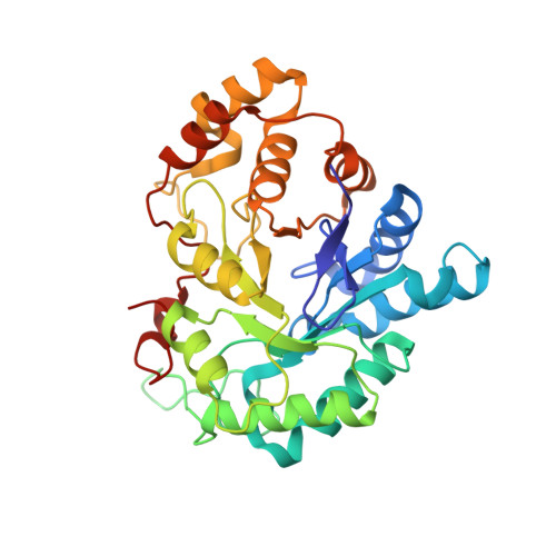
2DUX, 2DUZ, 2DV0, 2FZ8, 2FZ9, 2FZB, 2FZD, 2HV5, 2HVN, 2HVO - PubMed Abstract:
In structure-based drug design, accurate crystal structure determination of protein-ligand complexes is of utmost importance in order to elucidate the binding characteristics of a putative lead to a given target. It is the starting point for further design hypotheses to predict novel leads with improved properties. Often, crystal structure determination is regarded as ultimate proof for ligand binding providing detailed insight into the specific binding mode of the ligand to the protein. This widely accepted practise relies on the assumption that the crystal structure of a given protein-ligand complex is unique and independent of the protocol applied to produce the crystals. We present two examples indicating that this assumption is not generally given, even though the composition of the mother liquid for crystallisation was kept unchanged: Multiple crystal structure determinations of aldose reductase complexes obtained under varying crystallisation protocols concerning soaking and crystallisation exposure times were performed resulting in a total of 17 complete data sets and ten refined crystal structures, eight in complex with zopolrestat and two complexed with tolrestat. In the first example, a flip of a peptide bond is observed, obviously depending on the crystallisation protocol with respect to soaking and co-crystallisation conditions. This peptide flip is accompanied by a rupture of an H-bond formed to the bound ligand zopolrestat. The indicated enhanced local mobility of the complex is in agreement with the results of molecular dynamics simulations. As a second example, the aldose reductase-tolrestat complex is studied. Unexpectedly, two structures could be obtained: one with one, and a second with four inhibitor molecules bound to the protein. They are located in and near the binding pocket facilitated by crystal packing effects. Accommodation of the four ligand molecules is accompanied by pronounced shifts concerning two helices interacting with the additional ligands.
Organizational Affiliation:
Department of Pharmaceutical Chemistry, Philipps-University Marburg, Marbacher Weg 6, 35032 Marburg, Germany.