2F9Z

Complex between the chemotaxis deamidase CheD and the chemotaxis phosphatase CheC from Thermotoga maritima


Experimental Data Snapshot

  • Method: X-RAY DIFFRACTION
  • Resolution: 2.40 Å
  • R-Value Free: 0.275 
  • R-Value Work: 0.211 
  • R-Value Observed: 0.211 

wwPDB Validation   3D Report Full Report


This is version 1.4 of the entry. See complete history


Literature

A receptor-modifying deamidase in complex with a signaling phosphatase reveals reciprocal regulation.

Chao, X.Muff, T.J.Park, S.Y.Zhang, S.Pollard, A.M.Ordal, G.W.Bilwes, A.M.Crane, B.R.

(2006) Cell 124: 561-571

  • DOI: https://doi.org/10.1016/j.cell.2005.11.046
  • Primary Citation of Related Structures:  
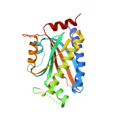
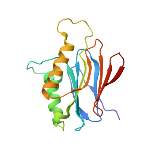
    2F9Z

  • PubMed Abstract: 

    Signal transduction underlying bacterial chemotaxis involves excitatory phosphorylation and feedback control through deamidation and methylation of sensory receptors. The structure of a complex between the signal-terminating phosphatase, CheC, and the receptor-modifying deamidase, CheD, reveals how CheC mimics receptor substrates to inhibit CheD and how CheD stimulates CheC phosphatase activity. CheD resembles other cysteine deamidases from bacterial pathogens that inactivate host Rho-GTPases. CheD not only deamidates receptor glutamine residues contained within a conserved structural motif but also hydrolyzes glutamyl-methyl-esters at select regulatory positions. Substituting Gln into the receptor motif of CheC turns the inhibitor into a CheD substrate. Phospho-CheY, the intracellular signal and CheC target, stabilizes the CheC:CheD complex and reduces availability of CheD. A point mutation that dissociates CheC from CheD impairs chemotaxis in vivo. Thus, CheC incorporates an element of an upstream receptor to influence both its own effect on receptor output and that of its binding partner, CheD.


  • Organizational Affiliation

    Department of Chemistry and Chemical Biology, Cornell University, Ithaca, NY 14853, USA.


Macromolecules
Find similar proteins by:  (by identity cutoff)  |  3D Structure
Entity ID: 1
MoleculeChains Sequence LengthOrganismDetailsImage
chemotaxis protein CheC
A, B
205Thermotoga maritima MSB8Mutation(s): 0 
Gene Names: TM0904
UniProt
Find proteins for Q9X006 (Thermotoga maritima (strain ATCC 43589 / DSM 3109 / JCM 10099 / NBRC 100826 / MSB8))
Explore Q9X006 
Go to UniProtKB:  Q9X006
Entity Groups  
Sequence Clusters30% Identity50% Identity70% Identity90% Identity95% Identity100% Identity
UniProt GroupQ9X006
Sequence Annotations
Expand
  • Reference Sequence
Find similar proteins by:  (by identity cutoff)  |  3D Structure
Entity ID: 2
MoleculeChains Sequence LengthOrganismDetailsImage
PROTEIN (chemotaxis methylation protein)
C, D
159Thermotoga maritima MSB8Mutation(s): 0 
Gene Names: TM0903
UniProt
Find proteins for Q9X005 (Thermotoga maritima (strain ATCC 43589 / DSM 3109 / JCM 10099 / NBRC 100826 / MSB8))
Explore Q9X005 
Go to UniProtKB:  Q9X005
Entity Groups  
Sequence Clusters30% Identity50% Identity70% Identity90% Identity95% Identity100% Identity
UniProt GroupQ9X005
Sequence Annotations
Expand
  • Reference Sequence
Experimental Data & Validation

Experimental Data

  • Method: X-RAY DIFFRACTION
  • Resolution: 2.40 Å
  • R-Value Free: 0.275 
  • R-Value Work: 0.211 
  • R-Value Observed: 0.211 
  • Space Group: P 32
Unit Cell:
Length ( Å )Angle ( ˚ )
a = 66.08α = 90
b = 66.08β = 90
c = 161.76γ = 120
Software Package:
Software NamePurpose
HKL-2000data collection
SCALEPACKdata scaling
AMoREphasing
CNSrefinement
HKL-2000data reduction

Structure Validation

View Full Validation Report


Entry History 

Deposition Data

Revision History  (Full details and data files)

  • Version 1.0: 2006-06-06
    Type: Initial release
  • Version 1.1: 2008-05-01
    Changes: Version format compliance
  • Version 1.2: 2011-07-13
    Changes: Source and taxonomy, Version format compliance
  • Version 1.3: 2018-01-24
    Changes: Structure summary
  • Version 1.4: 2023-08-30
    Changes: Data collection, Database references, Refinement description