2EXK

Structure of the family43 beta-Xylosidase E187G from geobacillus stearothermophilus in complex with xylobiose


Experimental Data Snapshot

  • Method: X-RAY DIFFRACTION
  • Resolution: 2.20 Å
  • R-Value Free: 0.238 
  • R-Value Work: 0.185 
  • R-Value Observed: 0.188 

wwPDB Validation   3D Report Full Report


This is version 2.2 of the entry. See complete history


Literature

The Structure of an Inverting GH43 beta-Xylosidase from Geobacillus stearothermophilus with its Substrate Reveals the Role of the Three Catalytic Residues.

Brux, C.Ben-David, A.Shallom-Shezifi, D.Leon, M.Niefind, K.Shoham, G.Shoham, Y.Schomburg, D.

(2006) J Mol Biol 359: 97-109

  • DOI: https://doi.org/10.1016/j.jmb.2006.03.005
  • Primary Citation of Related Structures:  
    2EXH, 2EXI, 2EXJ, 2EXK

  • PubMed Abstract: 

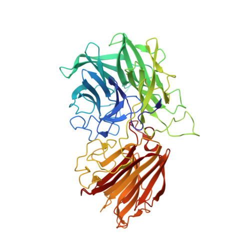
    beta-D-Xylosidases are glycoside hydrolases that catalyze the release of xylose units from short xylooligosaccharides and are engaged in the final breakdown of plant cell-wall hemicellulose. Here we describe the enzyme-substrate crystal structure of an inverting family 43 beta-xylosidase, from Geobacillus stearothermophilus T-6 (XynB3). Each XynB3 monomeric subunit is organized in two domains: an N-terminal five-bladed beta-propeller catalytic domain, and a beta-sandwich domain. The active site possesses a pocket topology, which is mainly constructed from the beta-propeller domain residues, and is closed on one side by a loop that originates from the beta-sandwich domain. This loop restricts the length of xylose units that can enter the active site, consistent with the exo mode of action of the enzyme. Structures of the enzyme-substrate (xylobiose) complex provide insights into the role of the three catalytic residues. The xylose moiety at the -1 subsite is held by a large number of hydrogen bonds, whereas only one hydroxyl of the xylose unit at the +1 subsite can create hydrogen bonds with the enzyme. The general base, Asp15, is located on the alpha-side of the -1 xylose sugar ring, 5.2 Angstroms from the anomeric carbon. This location enables it to activate a water molecule for a single-displacement attack on the anomeric carbon, resulting in inversion of the anomeric configuration. Glu187, the general acid, is 2.4 Angstroms from the glycosidic oxygen atom and can protonate the leaving aglycon. The third catalytic carboxylic acid, Asp128, is 4 Angstroms from the general acid; modulating its pK(a) and keeping it in the correct orientation relative to the substrate. In addition, Asp128 plays an important role in substrate binding via the 2-O of the glycon, which is important for the transition-state stabilization. Taken together, these key roles explain why Asp128 is an invariant among all five-bladed beta-propeller glycoside hydrolases.


  • Organizational Affiliation

    Institute for Biochemistry, University of Cologne, Germany.


Macromolecules
Find similar proteins by:  (by identity cutoff)  |  3D Structure
Entity ID: 1
MoleculeChains Sequence LengthOrganismDetailsImage
beta-D-xylosidase
A, B, C, D
535Geobacillus stearothermophilusMutation(s): 2 
EC: 3.2.1.37
UniProt
Find proteins for Q09LX0 (Geobacillus stearothermophilus)
Explore Q09LX0 
Go to UniProtKB:  Q09LX0
Entity Groups  
Sequence Clusters30% Identity50% Identity70% Identity90% Identity95% Identity100% Identity
UniProt GroupQ09LX0
Sequence Annotations
Expand
  • Reference Sequence
Oligosaccharides

Help

Entity ID: 2
MoleculeChains Length2D Diagram Glycosylation3D Interactions
alpha-D-xylopyranose-(1-4)-alpha-D-xylopyranose
E, F, G, H
2N/A
Glycosylation Resources
GlyTouCan:  G92719DV
GlyCosmos:  G92719DV
Small Molecules
Ligands 3 Unique
IDChains Name / Formula / InChI Key2D Diagram3D Interactions
MES
Query on MES

Download Ideal Coordinates CCD File 
J [auth A],
M [auth B],
R [auth D]
2-(N-MORPHOLINO)-ETHANESULFONIC ACID
C6 H13 N O4 S
SXGZJKUKBWWHRA-UHFFFAOYSA-N
GOL
Query on GOL

Download Ideal Coordinates CCD File 
K [auth A],
N [auth B],
P [auth C],
S [auth D]
GLYCEROL
C3 H8 O3
PEDCQBHIVMGVHV-UHFFFAOYSA-N
CA
Query on CA

Download Ideal Coordinates CCD File 
I [auth A],
L [auth B],
O [auth C],
Q [auth D]
CALCIUM ION
Ca
BHPQYMZQTOCNFJ-UHFFFAOYSA-N
Experimental Data & Validation

Experimental Data

  • Method: X-RAY DIFFRACTION
  • Resolution: 2.20 Å
  • R-Value Free: 0.238 
  • R-Value Work: 0.185 
  • R-Value Observed: 0.188 
  • Space Group: P 43 21 2
Unit Cell:
Length ( Å )Angle ( ˚ )
a = 139.87α = 90
b = 139.87β = 90
c = 232.025γ = 90
Software Package:
Software NamePurpose
DENZOdata reduction
SCALEPACKdata scaling
AMoREphasing
REFMACrefinement
PDB_EXTRACTdata extraction

Structure Validation

View Full Validation Report


Entry History 

Deposition Data

Revision History  (Full details and data files)

  • Version 1.0: 2006-04-04
    Type: Initial release
  • Version 1.1: 2008-05-01
    Changes: Version format compliance
  • Version 1.2: 2011-07-13
    Changes: Non-polymer description, Version format compliance
  • Version 1.3: 2017-10-18
    Changes: Advisory, Refinement description
  • Version 2.0: 2020-07-29
    Type: Remediation
    Reason: Carbohydrate remediation
    Changes: Advisory, Atomic model, Data collection, Derived calculations, Structure summary
  • Version 2.1: 2021-10-20
    Changes: Database references, Structure summary
  • Version 2.2: 2024-02-14
    Changes: Data collection