2EUD

Structures of Yeast Ribonucleotide Reductase I complexed with Ligands and Subunit Peptides


Experimental Data Snapshot

  • Method: X-RAY DIFFRACTION
  • Resolution: 2.30 Å
  • R-Value Free: 0.240 
  • R-Value Work: 0.204 
  • R-Value Observed: 0.210 

wwPDB Validation   3D Report Full Report


Ligand Structure Quality Assessment 


This is version 1.4 of the entry. See complete history


Literature

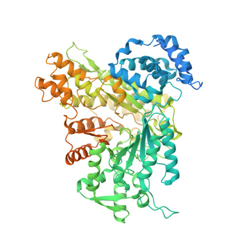
Structures of eukaryotic ribonucleotide reductase I define gemcitabine diphosphate binding and subunit assembly.

Xu, H.Faber, C.Uchiki, T.Racca, J.Dealwis, C.

(2006) Proc Natl Acad Sci U S A 103: 4028-4033

  • DOI: https://doi.org/10.1073/pnas.0600440103
  • Primary Citation of Related Structures:  
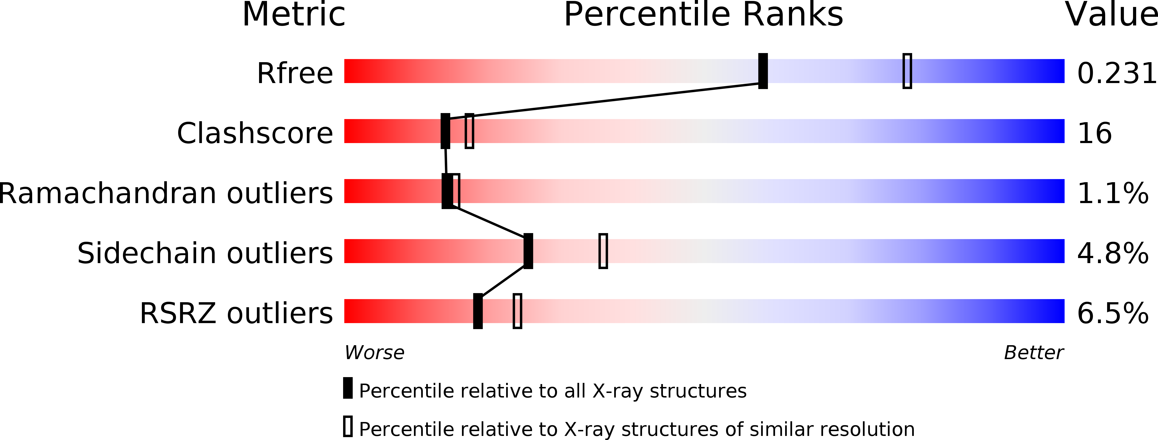
    2EUD

  • PubMed Abstract: 

    Ribonucleotide reductase (RNR) catalyzes the conversion of nucleoside diphosphates to deoxynucleoside diphosphates. Crucial for rapidly dividing cells, RNR is a target for cancer therapy. In eukaryotes, RNR comprises a heterooligomer of alpha(2) and beta(2) subunits. Rnr1, the alpha subunit, contains regulatory and catalytic sites; Rnr2, the beta subunit (in yeast, a heterodimer of Rnr2 and Rnr4), houses the diferric-tyrosyl radical crucial for catalysis. Here, we present three x-ray structures of eukaryotic Rnr1 from Saccharomyces cerevisiae: one bound to gemcitabine diphosphate (GemdP), the active metabolite of the mechanism-based chemotherapeutic agent gemcitabine; one with an Rnr2-derived peptide, and one with an Rnr4-derived peptide. Our structures reveal that GemdP binds differently from its analogue, cytidine diphosphate; because of unusual interactions of the geminal fluorines, the ribose and base of GemdP shift substantially, and loop 2, which mediates substrate specificity, adopts different conformations when binding to GemdP and cytidine diphosphate. The Rnr2 and Rnr4 peptides, which block RNR assembly, bind differently from each other but have unique modes of binding not seen in prokaryotic RNR. The Rnr2 peptide adopts a conformation similar to that previously reported from an NMR study for a mouse Rnr2-based peptide. In yeast, the Rnr2 peptide binds at subsites consisting of residues that are highly conserved among yeast, mouse, and human Rnr1s, suggesting that the mode of Rnr1-Rnr2 binding is conserved among eukaryotes. These structures provide new insights into subunit assembly and a framework for structure-based drug design targeting RNR.


  • Organizational Affiliation

    Department of Biochemistry and Cellular and Molecular Biology, University of Tennessee, M407 Walters Life Sciences, Knoxville, TN 37996-0840, USA.


Macromolecules
Find similar proteins by:  (by identity cutoff)  |  3D Structure
Entity ID: 1
MoleculeChains Sequence LengthOrganismDetailsImage
Ribonucleoside-diphosphate reductase large chain 1888Saccharomyces cerevisiaeMutation(s): 0 
Gene Names: RNR1
EC: 1.17.4.1
UniProt
Find proteins for P21524 (Saccharomyces cerevisiae (strain ATCC 204508 / S288c))
Explore P21524 
Go to UniProtKB:  P21524
Entity Groups  
Sequence Clusters30% Identity50% Identity70% Identity90% Identity95% Identity100% Identity
UniProt GroupP21524
Sequence Annotations
Expand
  • Reference Sequence
Small Molecules
Ligands 3 Unique
IDChains Name / Formula / InChI Key2D Diagram3D Interactions
ANP
Query on ANP

Download Ideal Coordinates CCD File 
C [auth A]PHOSPHOAMINOPHOSPHONIC ACID-ADENYLATE ESTER
C10 H17 N6 O12 P3
PVKSNHVPLWYQGJ-KQYNXXCUSA-N
GCQ
Query on GCQ

Download Ideal Coordinates CCD File 
D [auth A]GEMCITABINE DIPHOSPHATE
C9 H13 F2 N3 O10 P2
FRQISCZGNNXEMD-QPPQHZFASA-N
MG
Query on MG

Download Ideal Coordinates CCD File 
B [auth A]MAGNESIUM ION
Mg
JLVVSXFLKOJNIY-UHFFFAOYSA-N
Experimental Data & Validation

Experimental Data

  • Method: X-RAY DIFFRACTION
  • Resolution: 2.30 Å
  • R-Value Free: 0.240 
  • R-Value Work: 0.204 
  • R-Value Observed: 0.210 
  • Space Group: P 21 21 2
Unit Cell:
Length ( Å )Angle ( ˚ )
a = 107.578α = 90
b = 117.216β = 90
c = 63.99γ = 90
Software Package:
Software NamePurpose
HKL-2000data collection
SCALEPACKdata scaling
CNSrefinement
HKL-2000data reduction
CNSphasing

Structure Validation

View Full Validation Report


Ligand Structure Quality Assessment 


Entry History 

Deposition Data

Revision History  (Full details and data files)

  • Version 1.0: 2006-03-07
    Type: Initial release
  • Version 1.1: 2008-05-01
    Changes: Version format compliance
  • Version 1.2: 2011-07-13
    Changes: Version format compliance
  • Version 1.3: 2016-02-17
    Changes: Structure summary
  • Version 1.4: 2024-02-14
    Changes: Data collection, Database references, Derived calculations