


X-ray structure of hypothetical selenium-binding protein from Sulfolobus tokodaii, ST0059
Yamada, M., Yoshida, H., Kuramitsu, S., Kamitori, S.To be published.
Experimental Data Snapshot
wwPDB Validation 3D Report Full Report

Entity ID: 1 | |||||
|---|---|---|---|---|---|
| Molecule | Chains | Sequence Length | Organism | Details | Image |
| 462aa long hypothetical selenium-binding protein | 462 | Sulfurisphaera tokodaii | Mutation(s): 0 | 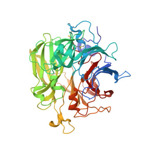 | |
UniProt | |||||
Find proteins for Q976Y0 (Sulfurisphaera tokodaii (strain DSM 16993 / JCM 10545 / NBRC 100140 / 7)) Explore Q976Y0 Go to UniProtKB: Q976Y0 | |||||
Entity Groups | |||||
| Sequence Clusters | 30% Identity50% Identity70% Identity90% Identity95% Identity100% Identity | ||||
| UniProt Group | Q976Y0 | ||||
Sequence AnnotationsExpand | |||||
| |||||
| Length ( Å ) | Angle ( ˚ ) |
|---|---|
| a = 142.196 | α = 90 |
| b = 142.196 | β = 90 |
| c = 142.196 | γ = 90 |
| Software Name | Purpose |
|---|---|
| CNS | refinement |
| ADSC | data collection |
| HKL-2000 | data reduction |
| HKL-2000 | data scaling |
| MLPHARE | phasing |
| CNS | phasing |
RCSB PDB (citation) is hosted by
RCSB PDB is a member of the

