Structural Insights into Unique Substrate Selectivity of Thermoplasma acidophilumd-Aldohexose Dehydrogenase
Yasutake, Y., Nishiya, Y., Tamura, N., Tamura, T.(2007) J Mol Biol 367: 1034-1046
- PubMed: 17300803
- DOI: https://doi.org/10.1016/j.jmb.2007.01.029
- Primary Citation of Related Structures:
2DTD, 2DTE, 2DTX - PubMed Abstract:
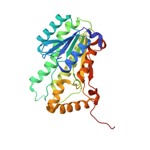
The D-aldohexose dehydrogenase from the thermoacidophilic archaea Thermoplasma acidophilum (AldT) belongs to the short-chain dehydrogenase/reductase (SDR) superfamily and catalyzes the oxidation of several monosaccharides with a preference for NAD(+) rather than NADP(+) as a cofactor. It has been found that AldT is a unique enzyme that exhibits the highest dehydrogenase activity against D-mannose. Here, we describe the crystal structures of AldT in ligand-free form, in complex with NADH, and in complex with the substrate D-mannose, at 2.1 A, 1.65 A, and 1.6 A resolution, respectively. The AldT subunit forms a typical SDR fold with an unexpectedly long C-terminal tail and assembles into an intertwined tetramer. The D-mannose complex structure reveals that Glu84 interacts with the axial C2 hydroxyl group of the bound D-mannose. Structural comparison with Bacillus megaterium glucose dehydrogenase (BmGlcDH) suggests that the conformation of the glutamate side-chain is crucial for discrimination between D-mannose and its C2 epimer D-glucose, and the conformation of the glutamate side-chain depends on the spatial arrangement of nearby hydrophobic residues that do not directly interact with the substrate. Elucidation of the D-mannose recognition mechanism of AldT further provides structural insights into the unique substrate selectivity of AldT. Finally, we show that the extended C-terminal tail completely shuts the substrate-binding pocket of the neighboring subunit both in the presence and absence of substrate. The elaborate inter-subunit interactions between the C-terminal tail and the entrance of the substrate-binding pocket imply that the tail may play a pivotal role in the enzyme activity.
Organizational Affiliation:
Research Institute of Genome-based Biofactory, National Institute of Advanced Industrial Science and Technology, Toyohira-ku, Sapporo 062-8517, Japan.