2DE5

Crystal structure of the electron transfer complex between oxygenase and ferredoxin in carbazole 1,9a-dioxygenase


Experimental Data Snapshot

  • Method: X-RAY DIFFRACTION
  • Resolution: 1.90 Å
  • R-Value Free: 0.233 
  • R-Value Work: 0.199 
  • R-Value Observed: 0.199 

wwPDB Validation   3D Report Full Report


This is version 1.3 of the entry. See complete history


Literature

Electron Transfer Complex Formation between Oxygenase and Ferredoxin Components in Rieske Nonheme Iron Oxygenase System

Ashikawa, Y.Fujimoto, Z.Noguchi, H.Habe, H.Omori, T.Yamane, H.Nojiri, H.

(2006) Structure 14: 1779-1789

  • DOI: https://doi.org/10.1016/j.str.2006.10.004
  • Primary Citation of Related Structures:  
    2DE5, 2DE6, 2DE7

  • PubMed Abstract: 

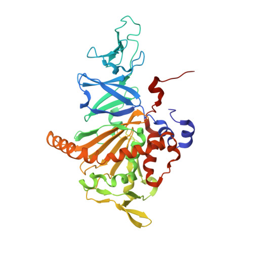
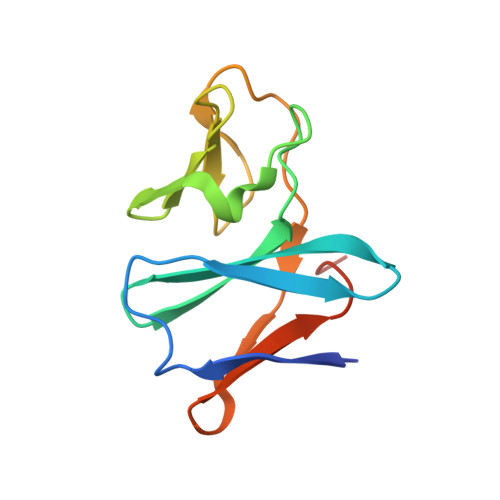
    Carbazole 1,9a-dioxygenase (CARDO), a member of the Rieske nonheme iron oxygenase system (ROS), consists of a terminal oxygenase (CARDO-O) and electron transfer components (ferredoxin [CARDO-F] and ferredoxin reductase [CARDO-R]). We determined the crystal structures of the nonreduced, reduced, and substrate-bound binary complexes of CARDO-O with its electron donor, CARDO-F, at 1.9, 1.8, and 2.0 A resolutions, respectively. These structures provide the first structure-based interpretation of intercomponent electron transfer between two Rieske [2Fe-2S] clusters of ferredoxin and oxygenase in ROS. Three molecules of CARDO-F bind to the subunit boundary of one CARDO-O trimeric molecule, and specific binding created by electrostatic and hydrophobic interactions with conformational changes suitably aligns the two Rieske clusters for electron transfer. Additionally, conformational changes upon binding carbazole resulted in the closure of a lid over the substrate-binding pocket, thereby seemingly trapping carbazole at the substrate-binding site.


  • Organizational Affiliation

    Biotechnology Research Center, The University of Tokyo, 1-1-1 Yayoi, Bunkyo-ku, Tokyo 113-8657, Japan.


Macromolecules
Find similar proteins by:  (by identity cutoff)  |  3D Structure
Entity ID: 1
MoleculeChains Sequence LengthOrganismDetailsImage
terminal oxygenase component of carbazole
A, B, C
392JanthinobacteriumMutation(s): 0 
EC: 1.14.12
UniProt
Find proteins for Q84II6 (Janthinobacterium sp. (strain J3))
Explore Q84II6 
Go to UniProtKB:  Q84II6
Entity Groups  
Sequence Clusters30% Identity50% Identity70% Identity90% Identity95% Identity100% Identity
UniProt GroupQ84II6
Sequence Annotations
Expand
  • Reference Sequence
Find similar proteins by:  (by identity cutoff)  |  3D Structure
Entity ID: 2
MoleculeChains Sequence LengthOrganismDetailsImage
ferredoxin component of carbazole
D, E, F
115Pseudomonas resinovoransMutation(s): 0 
EC: 1.14.12
UniProt
Find proteins for Q8GI16 (Pseudomonas resinovorans)
Explore Q8GI16 
Go to UniProtKB:  Q8GI16
Entity Groups  
Sequence Clusters30% Identity50% Identity70% Identity90% Identity95% Identity100% Identity
UniProt GroupQ8GI16
Sequence Annotations
Expand
  • Reference Sequence
Small Molecules
Experimental Data & Validation

Experimental Data

  • Method: X-RAY DIFFRACTION
  • Resolution: 1.90 Å
  • R-Value Free: 0.233 
  • R-Value Work: 0.199 
  • R-Value Observed: 0.199 
  • Space Group: P 1 21 1
Unit Cell:
Length ( Å )Angle ( ˚ )
a = 98.091α = 90
b = 89.835β = 103.79
c = 104.914γ = 90
Software Package:
Software NamePurpose
CNSrefinement
HKL-2000data reduction
SCALEPACKdata scaling
CNSphasing

Structure Validation

View Full Validation Report


Entry History 

Deposition Data

Revision History  (Full details and data files)

  • Version 1.0: 2006-12-05
    Type: Initial release
  • Version 1.1: 2008-04-30
    Changes: Version format compliance
  • Version 1.2: 2011-07-13
    Changes: Version format compliance
  • Version 1.3: 2023-10-25
    Changes: Data collection, Database references, Derived calculations, Refinement description