2DD7

A GFP-like protein from marine copepod, Chiridius poppei


Experimental Data Snapshot

  • Method: X-RAY DIFFRACTION
  • Resolution: 1.90 Å
  • R-Value Free: 0.232 
  • R-Value Work: 0.204 

wwPDB Validation   3D Report Full Report


Ligand Structure Quality Assessment 


This is version 1.2 of the entry. See complete history


Literature

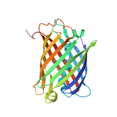
Structural basis for red-shifted emission of a GFP-like protein from the marine copepod Chiridius poppei

Suto, K.Masuda, H.Takenaka, Y.Tsuji, F.I.Mizuno, H.

(2009) Genes Cells 14: 727-737

  • DOI: https://doi.org/10.1111/j.1365-2443.2009.01305.x
  • Primary Citation of Related Structures:  
    2DD7, 2DD9

  • PubMed Abstract: 

    The fluorescence excitation and emission maxima of a GFP-like protein from the marine copepod Chiridius poppei (CpYGFP) show a significant red shift (lambda(ex) = 509 nm, lambda(em) = 517 nm) compared with those of GFP from Aequorea victoria (avGFP) and other GFP-like proteins from marine copepods. We performed crystallographic and biochemical studies to understand why this shift occurs in CpYGFP. The structure of CpYGFP showed that the imidazole side chain of His52 is involved in stacking on the phenol moiety of the chromophore. We investigated the potential role of His52 in causing the red-shifted spectral properties by performing mutational analyses of H52T, H52D and H52F. The emission wavelengths of H52T and H52D were blue-shifted and that of H52F was red-shifted relative to the wild type. Comparison of its structure of another copepod GFP (ppluGFP2) having an emission maximum at 502 nm showed that the imidazole ring of His54 (corresponding to His52 in CpYGFP) is flipped out of the stacking position with the chromophore. These findings suggest that pi-pi stacking interaction between His52 and the phenol moiety of the chromophore is the likely cause of the red-shift in light emission.


  • Organizational Affiliation

    VALWAY Technology Center, NEC Soft, Ltd, 1-18-7, Shinkiba, Koto-ku, Tokyo 136-8627, Japan.


Macromolecules
Find similar proteins by:  (by identity cutoff)  |  3D Structure
Entity ID: 1
MoleculeChains Sequence LengthOrganismDetailsImage
green fluorescent protein
A, B
216Chiridius poppeiMutation(s): 1 
UniProt
Find proteins for Q2MHN7 (Chiridius poppei)
Explore Q2MHN7 
Go to UniProtKB:  Q2MHN7
Entity Groups  
Sequence Clusters30% Identity50% Identity70% Identity90% Identity95% Identity100% Identity
UniProt GroupQ2MHN7
Sequence Annotations
Expand
  • Reference Sequence
Small Molecules
Ligands 2 Unique
IDChains Name / Formula / InChI Key2D Diagram3D Interactions
CXS
Query on CXS

Download Ideal Coordinates CCD File 
D [auth B]3-CYCLOHEXYL-1-PROPYLSULFONIC ACID
C9 H19 N O3 S
PJWWRFATQTVXHA-UHFFFAOYSA-N
CL
Query on CL

Download Ideal Coordinates CCD File 
C [auth A]CHLORIDE ION
Cl
VEXZGXHMUGYJMC-UHFFFAOYSA-M
Modified Residues  1 Unique
IDChains TypeFormula2D DiagramParent
CR2
Query on CR2
A, B
L-PEPTIDE LINKINGC13 H13 N3 O4GLY, TYR, GLY
Experimental Data & Validation

Experimental Data

  • Method: X-RAY DIFFRACTION
  • Resolution: 1.90 Å
  • R-Value Free: 0.232 
  • R-Value Work: 0.204 
  • Space Group: C 2 2 21
Unit Cell:
Length ( Å )Angle ( ˚ )
a = 113.478α = 90
b = 133.494β = 90
c = 108.733γ = 90
Software Package:
Software NamePurpose
CNSrefinement
MOSFLMdata reduction
CCP4data scaling
CNXphasing

Structure Validation

View Full Validation Report


Ligand Structure Quality Assessment 


Entry History 

Deposition Data

Revision History  (Full details and data files)

  • Version 1.0: 2007-01-23
    Type: Initial release
  • Version 1.1: 2008-04-30
    Changes: Version format compliance
  • Version 1.2: 2011-07-13
    Changes: Version format compliance