Crystal structure of TBP-interacting protein (Tk-TIP26) and implications for its inhibition mechanism of the interaction between TBP and TATA-DNA
Yamamoto, T., Matsuda, T., Inoue, T., Matsumura, H., Morikawa, M., Kanaya, S., Kai, Y.(2006) Protein Sci 15: 152-161
- PubMed: 16322571
- DOI: https://doi.org/10.1110/ps.051788906
- Primary Citation of Related Structures:
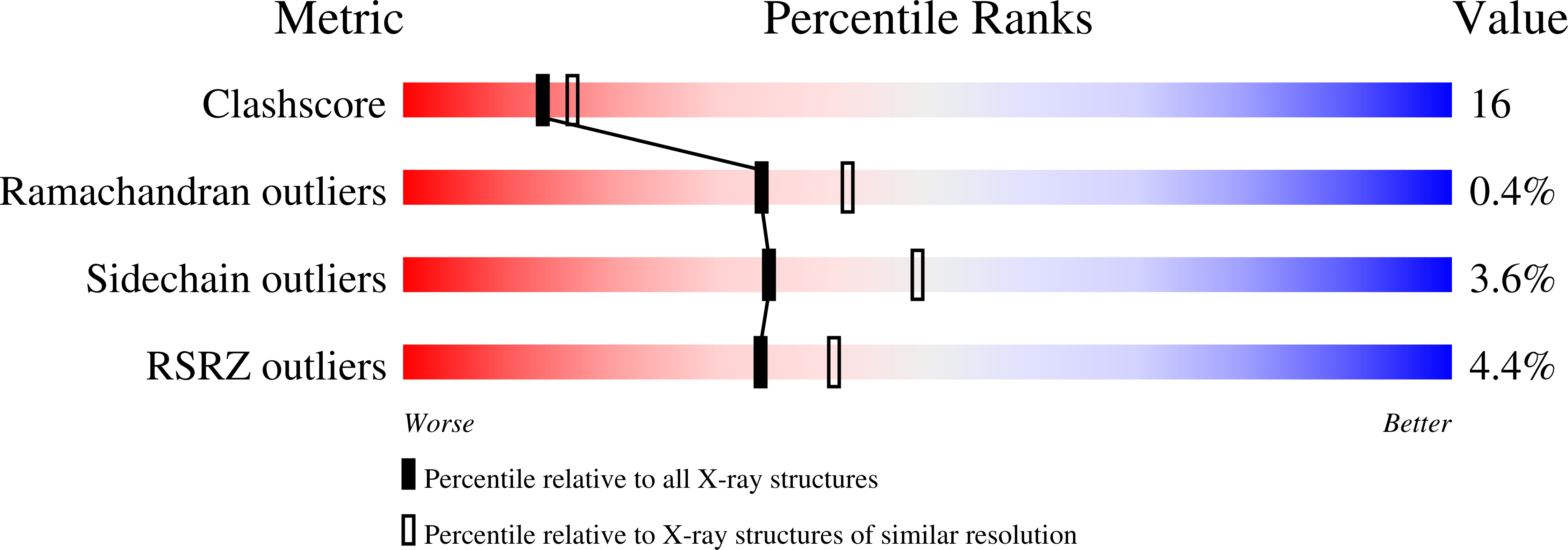
2CZR - PubMed Abstract:
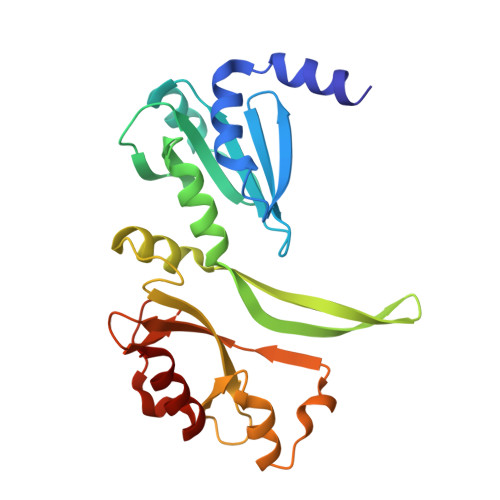
TATA-binding protein (TBP)-interacting protein from the hyperthermophilic archaeon Thermococcus kodakaraensis strain KOD1 (Tk-TIP26) is a possible transcription regulatory protein in Thermococcales. Here, we report the crystal structure of Tk-TIP26 determined at 2.3 A resolution with multiple-wavelength anomalous dispersion (MAD) method. The overall structure of Tk-TIP26 consists of two domains. The N-terminal domain forms an alpha/beta structure, in which three alpha-helices enclose the central beta-sheet. The topology of this domain is similar to that of holliday junction resolvase Hjc from Pyrococcus furiosus. The C-terminal domain comprises three alpha-helices, six beta-strands, and two 3(10)-helices. In the dimer structure of Tk-TIP26, two molecules are related with the crystallographic twofold axis, and these molecules rigidly interact with each other via hydrogen bonds. The complex of Tk-TIP26/Tk-TBP is isolated and analyzed by SDS-PAGE and gel filtration column chromatography, resulting in a stoichiometric ratio of the interaction between Tk-TIP26 and Tk-TBP of 4:2, i.e., two dimer molecules of Tk-TIP26 formed a complex with one dimeric TBP. The electrostatic surfaces of Tk-TIP26 and TBP from Pyrocuccus woesei (PwTBP) allowed us to build a model of the Tk-TIP26/TBP complex, and to propose the inhibition mechanism where two dimer molecules of Tk-TIP26 bind to a dimeric TBP, preventing its binding to TATA-DNA.
Organizational Affiliation:
Department of Applied Chemistry, Graduate School of Engineering, Osaka University, 2-1 Yamada-oka, Suita, Osaka 565-0871, Japan.