2CM8

Structural Basis for Inhibition of Protein Tyrosine Phosphatase 1B by Isothiazolidinone Heterocyclic Phosphonate Mimetics


Experimental Data Snapshot

  • Method: X-RAY DIFFRACTION
  • Resolution: 2.10 Å
  • R-Value Free: 0.253 
  • R-Value Work: 0.209 

wwPDB Validation   3D Report Full Report


This is version 1.3 of the entry. See complete history


Literature

Structural Basis for Inhibition of Protein-Tyrosine Phosphatase 1B by Isothiazolidinone Heterocyclic Phosphonate Mimetics.

Ala, P.J.Gonneville, L.Hillman, M.C.Becker-Pasha, M.Wei, M.Reid, B.G.Klabe, R.Yue, E.W.Wayland, B.Douty, B.Polam, P.Wasserman, Z.Bower, M.Combs, A.P.Burn, T.C.Hollis, G.F.Wynn, R.

(2006) J Biol Chem 281: 32784

  • DOI: https://doi.org/10.1074/jbc.M606873200
  • Primary Citation of Related Structures:  
    2CM2, 2CM3, 2CM7, 2CM8, 2CMA, 2CMB, 2CMC

  • PubMed Abstract: 

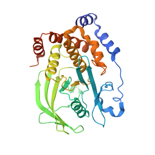
    Crystal structures of protein-tyrosine phosphatase 1B in complex with compounds bearing a novel isothiazolidinone (IZD) heterocyclic phosphonate mimetic reveal that the heterocycle is highly complementary to the catalytic pocket of the protein. The heterocycle participates in an extensive network of hydrogen bonds with the backbone of the phosphate-binding loop, Phe(182) of the flap, and the side chain of Arg(221). When substituted with a phenol, the small inhibitor induces the closed conformation of the protein and displaces all waters in the catalytic pocket. Saturated IZD-containing peptides are more potent inhibitors than unsaturated analogs because the IZD heterocycle and phenyl ring directly attached to it bind in a nearly orthogonal orientation with respect to each other, a conformation that is close to the energy minimum of the saturated IZD-phenyl moiety. These results explain why the heterocycle is a potent phosphonate mimetic and an ideal starting point for designing small nonpeptidic inhibitors.


  • Organizational Affiliation

    Incyte Corporation, Experimental Station, Route 141 and Henry Clay Road, Wilmington, DE 19880, USA. pauljala@gmail.com


Macromolecules
Find similar proteins by:  (by identity cutoff)  |  3D Structure
Entity ID: 1
MoleculeChains Sequence LengthOrganismDetailsImage
TYROSINE-PROTEIN PHOSPHATASE NON-RECEPTOR TYPE 1321Homo sapiensMutation(s): 0 
EC: 3.1.3.48
UniProt & NIH Common Fund Data Resources
Find proteins for P18031 (Homo sapiens)
Explore P18031 
Go to UniProtKB:  P18031
PHAROS:  P18031
GTEx:  ENSG00000196396 
Entity Groups  
Sequence Clusters30% Identity50% Identity70% Identity90% Identity95% Identity100% Identity
UniProt GroupP18031
Sequence Annotations
Expand
  • Reference Sequence
Small Molecules
Ligands 2 Unique
IDChains Name / Formula / InChI Key2D Diagram3D Interactions
F16
Query on F16

Download Ideal Coordinates CCD File 
B [auth A]5-(3-HYDROXYPHENYL)ISOTHIAZOL-3(2H)-ONE 1,1-DIOXIDE
C9 H7 N O4 S
FJABEDRGNJSWCC-UHFFFAOYSA-N
MG
Query on MG

Download Ideal Coordinates CCD File 
C [auth A]MAGNESIUM ION
Mg
JLVVSXFLKOJNIY-UHFFFAOYSA-N
Binding Affinity Annotations 
IDSourceBinding Affinity
F16 PDBBind:  2CM8 IC50: 1350 (nM) from 1 assay(s)
BindingDB:  2CM8 IC50: 1350 (nM) from 1 assay(s)
Binding MOAD:  2CM8 IC50: 1350 (nM) from 1 assay(s)
Experimental Data & Validation

Experimental Data

  • Method: X-RAY DIFFRACTION
  • Resolution: 2.10 Å
  • R-Value Free: 0.253 
  • R-Value Work: 0.209 
  • Space Group: P 31 2 1
Unit Cell:
Length ( Å )Angle ( ˚ )
a = 88.29α = 90
b = 88.29β = 90
c = 104.17γ = 120
Software Package:
Software NamePurpose
CNXrefinement
CrystalCleardata reduction
CrystalCleardata scaling
CNXphasing

Structure Validation

View Full Validation Report


Entry History 

Revision History  (Full details and data files)

  • Version 1.0: 2006-08-17
    Type: Initial release
  • Version 1.1: 2011-05-08
    Changes: Version format compliance
  • Version 1.2: 2011-07-13
    Changes: Version format compliance
  • Version 1.3: 2017-07-05
    Changes: Data collection