2CKL

Ring1b-Bmi1 E3 catalytic domain structure


Experimental Data Snapshot

  • Method: X-RAY DIFFRACTION
  • Resolution: 2.00 Å
  • R-Value Free: 0.213 
  • R-Value Work: 0.179 
  • R-Value Observed: 0.181 

wwPDB Validation   3D Report Full Report


This is version 1.1 of the entry. See complete history


Literature

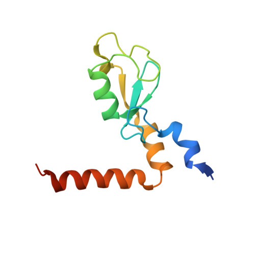
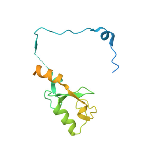
Structure and E3-Ligase Activity of the Ring-Ring Complex of Polycomb Proteins Bmi1 and Ring1B.

Buchwald, G.Van Der Stoop, P.Weichenrieder, O.Perrakis, A.Van Lohuizen, M.Sixma, T.K.

(2006) EMBO J 25: 2465

  • DOI: https://doi.org/10.1038/sj.emboj.7601144
  • Primary Citation of Related Structures:  
    2CKL

  • PubMed Abstract: 

    Polycomb group proteins Ring1b and Bmi1 (B-cell-specific Moloney murine leukaemia virus integration site 1) are critical components of the chromatin modulating PRC1 complex. Histone H2A ubiquitination by the PRC1 complex strongly depends on the Ring1b protein. Here we show that the E3-ligase activity of Ring1b on histone H2A is enhanced by Bmi1 in vitro. The N-terminal Ring-domains are sufficient for this activity and Ring1a can replace Ring1b. E2 enzymes UbcH5a, b, c or UbcH6 support this activity with varying processivity and selectivity. All four E2s promote autoubiquitination of Ring1b without affecting E3-ligase activity. We solved the crystal structure of the Ring-Ring heterodimeric complex of Ring1b and Bmi1. In the structure the arrangement of the Ring-domains is similar to another H2A E3 ligase, the BRCA1/BARD1 complex, but complex formation depends on an N-terminal arm of Ring1b that embraces the Bmi1 Ring-domain. Mutation of a critical residue in the E2/E3 interface shows that catalytic activity resides in Ring1b and not in Bmi1. These data provide a foundation for understanding the critical enzymatic activity at the core of the PRC1 polycomb complex, which is implicated in stem cell maintenance and cancer.


  • Organizational Affiliation

    Division of Molecular Carcinogenesis and Center for Biomedical Genetics, Netherlands Cancer Institute, Amsterdam, Netherlands.


Macromolecules
Find similar proteins by:  (by identity cutoff)  |  3D Structure
Entity ID: 1
MoleculeChains Sequence LengthOrganismDetailsImage
POLYCOMB GROUP RING FINGER PROTEIN 4108Mus musculusMutation(s): 0 
UniProt & NIH Common Fund Data Resources
Find proteins for P25916 (Mus musculus)
Explore P25916 
Go to UniProtKB:  P25916
IMPC:  MGI:88174
Entity Groups  
Sequence Clusters30% Identity50% Identity70% Identity90% Identity95% Identity100% Identity
UniProt GroupP25916
Sequence Annotations
Expand
  • Reference Sequence
Find similar proteins by:  (by identity cutoff)  |  3D Structure
Entity ID: 2
MoleculeChains Sequence LengthOrganismDetailsImage
UBIQUITIN LIGASE PROTEIN RING2165Mus musculusMutation(s): 0 
EC: 6.3.2
UniProt
Find proteins for Q9CQJ4 (Mus musculus)
Explore Q9CQJ4 
Go to UniProtKB:  Q9CQJ4
Entity Groups  
Sequence Clusters30% Identity50% Identity70% Identity90% Identity95% Identity100% Identity
UniProt GroupQ9CQJ4
Sequence Annotations
Expand
  • Reference Sequence
Experimental Data & Validation

Experimental Data

  • Method: X-RAY DIFFRACTION
  • Resolution: 2.00 Å
  • R-Value Free: 0.213 
  • R-Value Work: 0.179 
  • R-Value Observed: 0.181 
  • Space Group: P 63
Unit Cell:
Length ( Å )Angle ( ˚ )
a = 120.046α = 90
b = 120.046β = 90
c = 27.096γ = 120
Software Package:
Software NamePurpose
REFMACrefinement
MOSFLMdata reduction
SCALAdata scaling
SHARPphasing

Structure Validation

View Full Validation Report


Entry History 

Deposition Data

Revision History  (Full details and data files)

  • Version 1.0: 2006-05-11
    Type: Initial release
  • Version 1.1: 2011-07-13
    Changes: Advisory, Version format compliance