2C84

CRYSTAL STRUCTURE OF THE SIALYLTRANSFERASE PM0188 WITH CMP


Experimental Data Snapshot

  • Method: X-RAY DIFFRACTION
  • Resolution: 2.31 Å
  • R-Value Free: 0.227 
  • R-Value Work: 0.193 
  • R-Value Observed: 0.195 

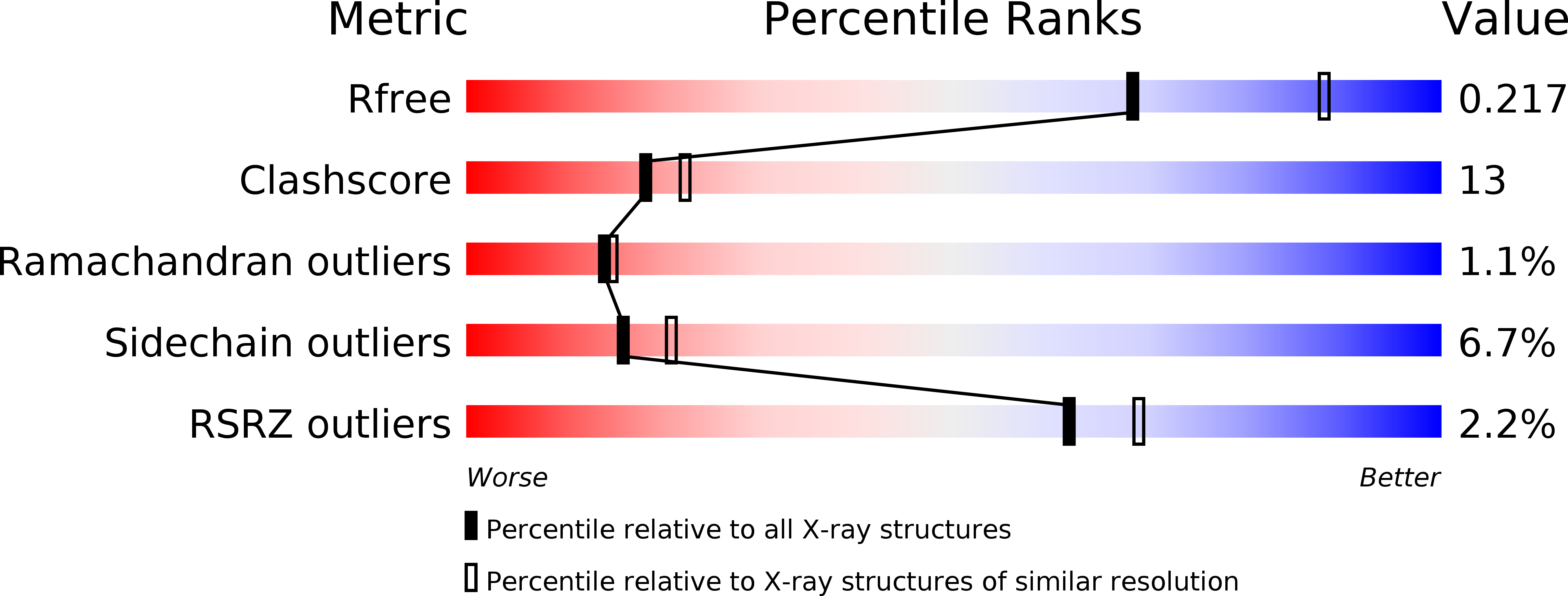
wwPDB Validation   3D Report Full Report


This is version 1.4 of the entry. See complete history


Literature

Structural analysis of sialyltransferase PM0188 from Pasteurella multocida complexed with donor analogue and acceptor sugar.

Kim, D.U.Yoo, J.H.Lee, Y.J.Kim, K.S.Cho, H.S.

(2008) BMB Rep 41: 48-54

  • DOI: https://doi.org/10.5483/bmbrep.2008.41.1.048
  • Primary Citation of Related Structures:  
    2C83, 2C84, 2IY7, 2IY8

  • PubMed Abstract: 

    PM0188 is a newly identified sialyltransferase from P. multocida which transfers sialic acid from cytidine 5'-monophosphonuraminic acid (CMP-NeuAc) to an acceptor sugar. Although sialyltransferases are involved in important biological functions like cell-cell recognition, cell differentiation and receptor-ligand interactions, little is known about their catalytic mechanism. Here, we report the X-ray crystal structures of PM0188 in the presence of an acceptor sugar and a donor sugar analogue, revealing the precise mechanism of sialic acid transfer. Site-directed mutagenesis, kinetic assays, and structural analysis show that Asp141, His311, Glu338, Ser355 and Ser356 are important catalytic residues; Asp141 is especially crucial as it acts as a general base. These complex structures provide insights into the mechanism of sialyltransferases and the structure-based design of specific inhibitors.


  • Organizational Affiliation

    Department of Biology and Protein Network Research Center, Yonsei University, Seoul, Korea.


Macromolecules
Find similar proteins by:  (by identity cutoff)  |  3D Structure
Entity ID: 1
MoleculeChains Sequence LengthOrganismDetailsImage
ALPHA-2,3/2,6-SIALYLTRANSFERASE/SIALIDASE389Pasteurella multocidaMutation(s): 0 
UniProt
Find proteins for Q15KI8 (Pasteurella multocida)
Explore Q15KI8 
Go to UniProtKB:  Q15KI8
Entity Groups  
Sequence Clusters30% Identity50% Identity70% Identity90% Identity95% Identity100% Identity
UniProt GroupQ15KI8
Sequence Annotations
Expand
  • Reference Sequence
Small Molecules
Ligands 1 Unique
IDChains Name / Formula / InChI Key2D Diagram3D Interactions
C
Query on C

Download Ideal Coordinates CCD File 
B [auth A]CYTIDINE-5'-MONOPHOSPHATE
C9 H14 N3 O8 P
IERHLVCPSMICTF-XVFCMESISA-N
Modified Residues  1 Unique
IDChains TypeFormula2D DiagramParent
MSE
Query on MSE
A
L-PEPTIDE LINKINGC5 H11 N O2 SeMET
Experimental Data & Validation

Experimental Data

  • Method: X-RAY DIFFRACTION
  • Resolution: 2.31 Å
  • R-Value Free: 0.227 
  • R-Value Work: 0.193 
  • R-Value Observed: 0.195 
  • Space Group: P 21 21 21
Unit Cell:
Length ( Å )Angle ( ˚ )
a = 65.712α = 90
b = 66.163β = 90
c = 101.94γ = 90
Software Package:
Software NamePurpose
REFMACrefinement
HKL-2000data scaling
MOLREPphasing

Structure Validation

View Full Validation Report


Entry History 

Deposition Data

Revision History  (Full details and data files)

  • Version 1.0: 2007-03-27
    Type: Initial release
  • Version 1.1: 2011-05-08
    Changes: Version format compliance
  • Version 1.2: 2011-07-13
    Changes: Version format compliance
  • Version 1.3: 2018-04-04
    Changes: Data collection, Source and taxonomy
  • Version 1.4: 2019-10-09
    Changes: Data collection, Database references, Derived calculations